Öz
Kaynakça
- Spinner RJ, Howe BM. Leg. In: Standring S, editor. Gray’s anatomy: the anatomical basis of clinical practice. 41st ed. New York (NY): Elsevier Limited; 2016. p. 1400–18.
- Naoum S. Should Hoffa’s fat pad be resected during total knee arthroplasty? A review of literature. Romanian Journal of Military Medicine 2022;125:196–201.
- Addison O, Marcus RL, Lastayo PC, Ryan AS. Intermuscular fat: a review of the consequences and causes. Int J Endocrinol 2014; 309570.
- Symonds ME. Adipose tissue biology. 2nd ed. New York (NY): Springer; 2017. p. 414.
- Vettor R, Milan G, Franzin C, Sanna M, De Coppi P, Rizzuto R, Federspil G. The origin of intermuscular adipose tissue and its pathophysiological implications. Am J Physiol Endocrinol Metab 2009;297:E987–8.
- Contreras-Shannon V, Ochoa O, Reyes-Reyna SM, Sun D, Michalek JE, Kuziel WA, McManus LM, Shireman PK. Fat accumulation with altered inflammation and regeneration in skeletal muscle of CCR2−/− mice following ischemic injury. Am J Physiol 2007;292:C953–67.
- Manini TM, Clark BC, Nalls MA, Goodpaster BH, Ploutz-Snyder LL, Harris TB. Reduced physical activity increases intermuscular adipose tissue in healthy young adults. Am J Clin Nutr 2007;85:377–84.
- Tuttle LJ, Sinacore DR, Mueller MJ. Intermuscular adipose tissue is muscle specific and associated with poor functional performance. J Aging Res 2012:1–7.
- Katsiaras A, Newman AB, Kriska A, Brach J, Krishnaswami S, Feingold E, Kritchevsky SB, Li R, Harris TB, Schwartz A, Goodpaster BH. Skeletal muscle fatigue, strength, and quality in the elderly: the health ABC study. J Appl Physiol 2005;99:210–6.
- Goodpaster BH, Carlson CL, Visser M, Kelley DE, Scherzinger A, Harris TB, Stamm E, Newman AB. Attenuation of skeletal muscle and strength in the elderly: the health ABC study. J Appl Physiol 2001;90:2157–65.
- Visser M, Goodpaster BH, Kritchevsky SB, Newman AB, Nevitt M, Rubin SM, Simonsick EM, Harris TB. Muscle mass, muscle strength, and muscle fat infiltration as predictors of incident mobility limitations in well-functioning older persons. J Gerontol A Biol Sci Med Sci 2005;60:324–33.
- Visser M, Kritchevsky SB, Goodpaster BH, Newman AB, Nevitt M, Stamm E, Harris TB. Leg muscle mass and composition in relation to lower extremity performance in men and women aged 70 to 79: the health, aging and body composition study. J Am Geriatr Soc 2002;50:897–904.
- Fontanella CG, Carniel EL, Frigo A, Macchi V, Porzionato A, Sarasin G, Rossato M, de Caro R, Natali AN. Investigation of biomechanical response of Hoffa’s fat pad and comparative characterization. J Mech Behav Biomed Mater 2017;67:1–9.
- Rosen ED, Spiegelman BM. What we talk about when we talk about fat. Cell 2014;156:20–44.
- Ghazzawi A, Theobald P, Pugh N, Byrne C, Nokes L. Quantifying the motion of Kager’s fat pad. J Orthop Rel Res 2009;27:1457–60.
- Theobald P, Bydder G, Dent C, Nokes L, Pugh N, Benjamin M. The functional anatomy of Kager’s fat pad in relation to retrocalcaneal problems and other hindfoot disorders. J Anat 2006;208:91–7.
- Shaw HM, Benjamin M. Structure–function relationships of entheses in relation to mechanical load and exercise. Scand J Med Sci Sports 2007;17:303–15.
- Fontanella CG, Macchi V, Carniel EL, Frigo A, Porzionato A, Picardi Edoardo EE, Favero M, Ruggieri P, de Caro R, Natali AN. Biomechanical behavior of Hoffa’s fat pad in healthy and osteoarthritic conditions: histological and mechanical investigations. Australas Phys Eng Sci Med 2018;41:657–67.
- Gallagher J, Tierney P, Murray P, O’Brien M. The infrapatellar fat pad: anatomy and clinical correlations. Knee Surg Sports Traumatol Arthrosc 2005;13:268–72.
- Vahlensieck M, Linneborn G, Schild H, Schmidt HM. Hoffa’s recess: incidence, morphology and differential diagnosis of the globular-shaped cleft in the infrapatellar fat pad of the knee on MRI and cadaver dissections. Eur Radiol 2002;12:90–3.
- Takumi O, Hirofumi T, Hiroshi A, Hiroki Y, Toshihiro M, Masatomo M, Takuma H, Tsukasa K. Presence of adipose tissue along the posteromedial tibial border. J Exp Orthop 2021;8:1–9.
- Ortiz-Miguel S, Miguel-Pérez M, Blasi J, Pérez-Bellmunt A, Ortiz-Sagristà JC, Möller I, Agullo JL, Iglesias P, Martinoli C. Compartments of the crural fascia: clinically relevant ultrasound, anatomical and histological findings. Surg Radiol Anat 2023;1603–17.
- Stecco C. Functional atlas of human fascial system. London, Edinburg: Elsevier Churchill Livingstone; 2015. p. 256.
- Fairclough J, Hayashi K, Toumi H, Lyons K, Bydder G, Phillips N, Best TM, Benjamin M. The functional anatomy of the iliotibial band during flexion and extension of the knee: implications for understanding iliotibial band syndrome. J Anat 2006;208:309–16.
- Döner RS, Keleş P, Karip B. Examination of anatomic and morphometric features of Kager’s triangle. Turkish Journal of Health Science and Life 2022;5:207–13.
- Christov C, Chrétien F, Abou-Khalil R, Bassez G, Vallet G, Authier FJ, Bassaglia Y, Shinin V, Tajbakhsh S, Chazaud B, Gherardi RK. Muscle satellite cells and endothelial cells: close neighbors and privileged partners. Mol Biol Cell 2007;18:1397–409.
- Clavert P, Dosch JC, Wolfram-Gabel R, Kahn JL. New findings on intermetacarpal fat pads: anatomy and imaging. Surg Radiol Anat 2006;28:351–4.
- Benjamin M, Redman S, Milz S, Büttner A, Amin A, Moriggl B, Brenner E, Emery P, McGonagle D, Bydder G. Adipose tissue at entheses: the rheumatological implications of its distribution. A potential site of pain and stress dissipation? Ann Rheum Dis 2004; 63:1549–55.
- Kesilmiş İ, Kurtoğlu Olgunus Z. Could the biomechanical characteristics of the crural fascia in the anterior compartment be related to the levels of anterior leg pain occur in athletes? Acta Medica 2023; 54(Supplement 2):45.
- Migliorini S. Triathlon medicine. Cham, Switzerland: Springer Nature Switzerland; 2020, p. 415.
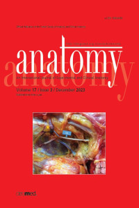
Morphometric description of the subfascial intermuscular adipose tissue of anterior compartment of the leg
Öz
Objectives: To describe the morphological characteristics of subfascial intermuscular adipose tissue (IMATS) in the anterior compartment of the leg, considering its developmental and functional relationship with the crural fascia.
Methods: In twenty formalin-fixed cadaveric legs (13 males, 7 females), after removal of the skin and crural fascia, the IMATS was exposed and classified into four types according to its shape. Leg length was divided into eight regions. The length, width at the widest point, closest distances of the upper and lower ends to the intermalleolar line and the anterior margin of the tibia, as well as the thickness of the skin-subcutaneous tissue complex, limb and leg lengths were measured for IMATS.
Results: The most common type of IMATS was the short-large type. The largest point of IMATS was located in zone 3 or 4, and this point was located in the two zones closest to the lower end of IMATS in 75% of cases. In all cases, one to three connecting vessels piercing the crural fascia (80% were in zones 2, 3 or 4) connected to the IMATS in a slightly lateral to medial oblique course of the IMATS from top to bottom. The IMATS was superficially located in the tendinous and muscular parts of the extensor digitorum longus and/or tibialis anterior muscles, loosely attached to the muscles and crural fascia, but not between the muscle fibers. Although the largest point (p=0.041) and the distance from the distal end to the anterior margin of the tibia were found to be greater in males (p=0.049), the gender difference disappeared when normalized for limb length.
Conclusion: No data on IMATS morphometry could be found in the literature. A remarkable finding of the study, which is open to interpretation in terms of the function of the IMATS, is that the location of the IMATS overlaps with the crural fascia region, which is reported to be biomechanically stiffer in the transverse direction. Our data that a connecting vessel is always connected to the IMATS by a fixed spatial relationship strengthens the argument that the developmental history of both structures may intersect.
Anahtar Kelimeler
anterior compartment of leg crural fascia extensor digitorum longus fat pad intermuscular adipose tissue tibialis anterior muscle
Kaynakça
- Spinner RJ, Howe BM. Leg. In: Standring S, editor. Gray’s anatomy: the anatomical basis of clinical practice. 41st ed. New York (NY): Elsevier Limited; 2016. p. 1400–18.
- Naoum S. Should Hoffa’s fat pad be resected during total knee arthroplasty? A review of literature. Romanian Journal of Military Medicine 2022;125:196–201.
- Addison O, Marcus RL, Lastayo PC, Ryan AS. Intermuscular fat: a review of the consequences and causes. Int J Endocrinol 2014; 309570.
- Symonds ME. Adipose tissue biology. 2nd ed. New York (NY): Springer; 2017. p. 414.
- Vettor R, Milan G, Franzin C, Sanna M, De Coppi P, Rizzuto R, Federspil G. The origin of intermuscular adipose tissue and its pathophysiological implications. Am J Physiol Endocrinol Metab 2009;297:E987–8.
- Contreras-Shannon V, Ochoa O, Reyes-Reyna SM, Sun D, Michalek JE, Kuziel WA, McManus LM, Shireman PK. Fat accumulation with altered inflammation and regeneration in skeletal muscle of CCR2−/− mice following ischemic injury. Am J Physiol 2007;292:C953–67.
- Manini TM, Clark BC, Nalls MA, Goodpaster BH, Ploutz-Snyder LL, Harris TB. Reduced physical activity increases intermuscular adipose tissue in healthy young adults. Am J Clin Nutr 2007;85:377–84.
- Tuttle LJ, Sinacore DR, Mueller MJ. Intermuscular adipose tissue is muscle specific and associated with poor functional performance. J Aging Res 2012:1–7.
- Katsiaras A, Newman AB, Kriska A, Brach J, Krishnaswami S, Feingold E, Kritchevsky SB, Li R, Harris TB, Schwartz A, Goodpaster BH. Skeletal muscle fatigue, strength, and quality in the elderly: the health ABC study. J Appl Physiol 2005;99:210–6.
- Goodpaster BH, Carlson CL, Visser M, Kelley DE, Scherzinger A, Harris TB, Stamm E, Newman AB. Attenuation of skeletal muscle and strength in the elderly: the health ABC study. J Appl Physiol 2001;90:2157–65.
- Visser M, Goodpaster BH, Kritchevsky SB, Newman AB, Nevitt M, Rubin SM, Simonsick EM, Harris TB. Muscle mass, muscle strength, and muscle fat infiltration as predictors of incident mobility limitations in well-functioning older persons. J Gerontol A Biol Sci Med Sci 2005;60:324–33.
- Visser M, Kritchevsky SB, Goodpaster BH, Newman AB, Nevitt M, Stamm E, Harris TB. Leg muscle mass and composition in relation to lower extremity performance in men and women aged 70 to 79: the health, aging and body composition study. J Am Geriatr Soc 2002;50:897–904.
- Fontanella CG, Carniel EL, Frigo A, Macchi V, Porzionato A, Sarasin G, Rossato M, de Caro R, Natali AN. Investigation of biomechanical response of Hoffa’s fat pad and comparative characterization. J Mech Behav Biomed Mater 2017;67:1–9.
- Rosen ED, Spiegelman BM. What we talk about when we talk about fat. Cell 2014;156:20–44.
- Ghazzawi A, Theobald P, Pugh N, Byrne C, Nokes L. Quantifying the motion of Kager’s fat pad. J Orthop Rel Res 2009;27:1457–60.
- Theobald P, Bydder G, Dent C, Nokes L, Pugh N, Benjamin M. The functional anatomy of Kager’s fat pad in relation to retrocalcaneal problems and other hindfoot disorders. J Anat 2006;208:91–7.
- Shaw HM, Benjamin M. Structure–function relationships of entheses in relation to mechanical load and exercise. Scand J Med Sci Sports 2007;17:303–15.
- Fontanella CG, Macchi V, Carniel EL, Frigo A, Porzionato A, Picardi Edoardo EE, Favero M, Ruggieri P, de Caro R, Natali AN. Biomechanical behavior of Hoffa’s fat pad in healthy and osteoarthritic conditions: histological and mechanical investigations. Australas Phys Eng Sci Med 2018;41:657–67.
- Gallagher J, Tierney P, Murray P, O’Brien M. The infrapatellar fat pad: anatomy and clinical correlations. Knee Surg Sports Traumatol Arthrosc 2005;13:268–72.
- Vahlensieck M, Linneborn G, Schild H, Schmidt HM. Hoffa’s recess: incidence, morphology and differential diagnosis of the globular-shaped cleft in the infrapatellar fat pad of the knee on MRI and cadaver dissections. Eur Radiol 2002;12:90–3.
- Takumi O, Hirofumi T, Hiroshi A, Hiroki Y, Toshihiro M, Masatomo M, Takuma H, Tsukasa K. Presence of adipose tissue along the posteromedial tibial border. J Exp Orthop 2021;8:1–9.
- Ortiz-Miguel S, Miguel-Pérez M, Blasi J, Pérez-Bellmunt A, Ortiz-Sagristà JC, Möller I, Agullo JL, Iglesias P, Martinoli C. Compartments of the crural fascia: clinically relevant ultrasound, anatomical and histological findings. Surg Radiol Anat 2023;1603–17.
- Stecco C. Functional atlas of human fascial system. London, Edinburg: Elsevier Churchill Livingstone; 2015. p. 256.
- Fairclough J, Hayashi K, Toumi H, Lyons K, Bydder G, Phillips N, Best TM, Benjamin M. The functional anatomy of the iliotibial band during flexion and extension of the knee: implications for understanding iliotibial band syndrome. J Anat 2006;208:309–16.
- Döner RS, Keleş P, Karip B. Examination of anatomic and morphometric features of Kager’s triangle. Turkish Journal of Health Science and Life 2022;5:207–13.
- Christov C, Chrétien F, Abou-Khalil R, Bassez G, Vallet G, Authier FJ, Bassaglia Y, Shinin V, Tajbakhsh S, Chazaud B, Gherardi RK. Muscle satellite cells and endothelial cells: close neighbors and privileged partners. Mol Biol Cell 2007;18:1397–409.
- Clavert P, Dosch JC, Wolfram-Gabel R, Kahn JL. New findings on intermetacarpal fat pads: anatomy and imaging. Surg Radiol Anat 2006;28:351–4.
- Benjamin M, Redman S, Milz S, Büttner A, Amin A, Moriggl B, Brenner E, Emery P, McGonagle D, Bydder G. Adipose tissue at entheses: the rheumatological implications of its distribution. A potential site of pain and stress dissipation? Ann Rheum Dis 2004; 63:1549–55.
- Kesilmiş İ, Kurtoğlu Olgunus Z. Could the biomechanical characteristics of the crural fascia in the anterior compartment be related to the levels of anterior leg pain occur in athletes? Acta Medica 2023; 54(Supplement 2):45.
- Migliorini S. Triathlon medicine. Cham, Switzerland: Springer Nature Switzerland; 2020, p. 415.
Ayrıntılar
| Birincil Dil | İngilizce |
|---|---|
| Konular | Plastik, Rekonstrüktif ve Estetik Cerrahi |
| Bölüm | Original Articles |
| Yazarlar | |
| Yayımlanma Tarihi | 29 Aralık 2023 |
| Yayımlandığı Sayı | Yıl 2023 Cilt: 17 Sayı: 3 |
Kaynak Göster
Anatomy is the official publication of the Turkish Society of Anatomy and Clinical Anatomy(TSACA).


