Abstract
References
- Arıncı K, Elhan A. Anatomi 1. cilt: Kemikler, eklemler, kaslar, iç organlar. Ankara: Güneş Tıp Kitabevleri; 2016. p. 62–3.
- Standring S. Gray’s anatomy: the anatomical basis of clinical practice: 42nd ed. China: Elsevier Health Sciences; 2021. p. 831–34.
- Karabulut AK, Köylüoğlu B, Uysal İ. Human foetal sacral length measurement for the assessment of foetal growth and development by ultrasonography and dissection. Anat Histol Embryol 2001;30:141–6.
- Liguoro D, Viejo-Fuertes D, Midy D, Guerin J. The posterior sacral foramina: an anatomical study. J Anat 1999;195:301–4.
- Povo A, Arantes M, Matzel K, Barbosa J, Ferreira M, Pais D, Rodríguez-Baeza A. Surface anatomical landmarks for the location of posterior sacral foramina in sacral nerve stimulation. Tech Colo-proctol 2016;20:859–64.
- Senoglu N, Senoglu M, Oksuz H, Gumusalan Y, Yuksel K, Zencirci B, Ezberci M, Kızılkanat E. Landmarks of the sacral hiatus for caudal epidural block: an anatomical study. Br J Anaesth 2005;95:692–5.
- Chen CP, Tang SF, Hsu T-C, Tsai W-C, Liu H-P, Chen MJ, Date E, Lew HL. Ultrasound guidance in caudal epidural needle placement. Anesthesiology 2004;101:181–4.
- Moore KL, Dalley AF, Agur AMR. Clinically oriented anatomy. 6th ed. Baltimore: Wolters Kluwer; Lippincott Williams & Wilkins; 2014. p. 451.
- Aggarwal A, Aggarwal A, Harjeet, Sahni D. Morphometry of sacral hiatus and its clinical relevance in caudal epidural block. Surg Radiol Anat 2009;31:793–800.
- Bagheri H, Govsa F. Anatomy of the sacral hiatus and its clinical relevance in caudal epidural block. Surg Radiol Anat 2017;39:943–51.
- Kilinç CY. Which technique should be choosed in posterior pelvic ring injuries: percutaneous sacroiliac screw fixation technique or posterior percutaneous transiliac plating technique? [Article in Turkish] Kırıkkale Üniversitesi Tıp Fakültesi Dergisi 2019;21:80–4.
- Basaloglu H, Turgut M, Taşer FA, Ceylan T, Başaloğlu HK, Ceylan AA. Morphometry of the sacrum for clinical use. Surg Radiol Anat 2005;27:467–71.
- Öğrenci A, Yaman O. Spinopelvic instrumentation techniques. [Article in Turkish] Türkiye Klinikleri Nöroşirürji - Özel Konular 2017;7:334–9.
- Yilmaz S, Tokpinar A, Aycan K, Tutkun RT, Kanter AG, Çimen K, Sümeyye U, Susar H. Morphometric evaluation of the sacrum. [Article in Turkish] Bozok Medical Journal 2018;8:13–7.
- Elvan Ö, ORS AB, Uzmansel D. Morphologic evaluation of dorsal surface of sacrum. [Article in Turkish] Mersin Üniversitesi Sağlık Bilimleri Dergisi 2021;14:87–95.
- Polat S, Kabakci A, Oksüzler FY, Oksüzler M, Yücel AH. Morphometric analysis and clinical anatomy of sacrum and sacral hiatus in Turkish healthy adults. Cukurova Medical Journal 2020;45:672–9.
- Singh A, Gupta R, Singh A. Morphological and morphometrical study of sacral hiatus of human sacrum. Natl J Integr Res Med 2018;9:65–73.
- Sinha Manisha B, Mrithunjay R, Soumitra T, Siddiqui A. Morphometry of first pedicle of sacrum and its clinical relevance. International Journal of Healthcare and Biomedical Research 2013;1:234–40.
- Morales-Ávalos R, Leyva-Villegas JI, Vílchez-Cavazos F, de León ÁRM-P, Elizondo-Omaña RE, Guzmán-López S. Morphometric characteristics of the sacrum in Mexican population. Its importance in lumbosacral fusion and fixation procedures. [Article in Spanish] Cir Cir 2012;80:528–35.
- Arman C, Naderi S, Kiray A, Aksu FT, Yılmaz HS, Tetik S, Korman E. The human sacrum and safe approaches for screw placement. J Clin Neurosci 2009;16:1046–9.
- Hassanein GH. Metric study of Egyptian sacrum for lumbo‐sacral fixation procedures. Clin Anat 2011;24:218–24.
- Nadeem G. Importance of knowing the level of sacral hiatus for caudal epidural anesthesia. Journal of Morphological Sciences 2014;31:9–13.
- Vasuki A, Sundaram KK, Nirmaladevi M, Jamuna M, Hebzibah DJ, Fenn T. Anatomical study of sacrum and its clinical significance. Annals of International Medical and Dental Research 2016;2:123–7.
- Malarvani T, Ganesh E, Nirmala P. Study of sacral hiatus in dry human sacra in Nepal, Parsa Region. International Journal of Anatomy and Research 2015;3:848–55.
- David SJ. Morphometric measurements of sacral hiatus in South Indian dry human sacra for safe caudal epidural block. International Journal of Anatomy and Research 2019;7:6911–7.
- Kujur B, Gaikwad MR. A study of variations in sacral hiatus and its clinical significance. International Journal of Interdisciplinary and Multidisciplinary Studies 2017;4:204–12.
- Nastoulis E, Karakasi M-V, Pavlidis P, Thomaidis V, Fiska A. Anatomy and clinical significance of sacral variations: a systematic review. Folia Morphol (Warsz) 2019;78:651–67.
- Kubavat DM, Nagar SK, Lakhani C, Ruparelia SS, Patel S, Varlekar P. A study of sacrum with three pairs of sacral foramina in Western India. Int J Med Sci Public Health 2012;1:127–31.
Abstract
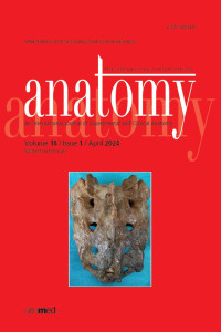
Objectives: The sacrum is a critical bone in various clinical procedures, including caudal epidural blocks, iliosacral screw placements, fetal growth assessment, and sacral nerve stimulation. This study aims to investigate the morphometry and variational morphology of the anatomical formations in the pelvis and dorsal surface of the sacrum.
Methods: Morphometric and morphological characteristics of 30 sacral bones of unknown age and sex were examined. Measurements were made using a digital caliper.
Results: The mean height of the sacrum was 106.67±10.16 mm, while their mean width was 103.60±6.78 mm. The morphometric analysis revealed that the mean length of the sacral hiatus was 18.51±7.44 mm, and the distance between the sacral cornua was 11.80±2.46 mm. The sacral hiatus was most commonly observed in an inverted ‘U’ shape, while the least common form was bifid. The sacral canal typically displayed a V-shaped morphology. It was determined that the apex of the sacral hiatus most frequently started at the S4 level (80%) compared to the sacral vertebra, and the base of the sacral hiatus mostly ended at the S5 level (93.4%).
Conclusion: Morphometry of the sacrum is essential in guiding clinicians, especially in interventions such as anesthesia and orthopedics. Discrepancies in parameter studies conducted in some countries suggest the significance of ethnic factors. Therefore, it is essential for the number of studies to increase and for physicians to follow the parameters of their society regarding the effectiveness of the treatments.
References
- Arıncı K, Elhan A. Anatomi 1. cilt: Kemikler, eklemler, kaslar, iç organlar. Ankara: Güneş Tıp Kitabevleri; 2016. p. 62–3.
- Standring S. Gray’s anatomy: the anatomical basis of clinical practice: 42nd ed. China: Elsevier Health Sciences; 2021. p. 831–34.
- Karabulut AK, Köylüoğlu B, Uysal İ. Human foetal sacral length measurement for the assessment of foetal growth and development by ultrasonography and dissection. Anat Histol Embryol 2001;30:141–6.
- Liguoro D, Viejo-Fuertes D, Midy D, Guerin J. The posterior sacral foramina: an anatomical study. J Anat 1999;195:301–4.
- Povo A, Arantes M, Matzel K, Barbosa J, Ferreira M, Pais D, Rodríguez-Baeza A. Surface anatomical landmarks for the location of posterior sacral foramina in sacral nerve stimulation. Tech Colo-proctol 2016;20:859–64.
- Senoglu N, Senoglu M, Oksuz H, Gumusalan Y, Yuksel K, Zencirci B, Ezberci M, Kızılkanat E. Landmarks of the sacral hiatus for caudal epidural block: an anatomical study. Br J Anaesth 2005;95:692–5.
- Chen CP, Tang SF, Hsu T-C, Tsai W-C, Liu H-P, Chen MJ, Date E, Lew HL. Ultrasound guidance in caudal epidural needle placement. Anesthesiology 2004;101:181–4.
- Moore KL, Dalley AF, Agur AMR. Clinically oriented anatomy. 6th ed. Baltimore: Wolters Kluwer; Lippincott Williams & Wilkins; 2014. p. 451.
- Aggarwal A, Aggarwal A, Harjeet, Sahni D. Morphometry of sacral hiatus and its clinical relevance in caudal epidural block. Surg Radiol Anat 2009;31:793–800.
- Bagheri H, Govsa F. Anatomy of the sacral hiatus and its clinical relevance in caudal epidural block. Surg Radiol Anat 2017;39:943–51.
- Kilinç CY. Which technique should be choosed in posterior pelvic ring injuries: percutaneous sacroiliac screw fixation technique or posterior percutaneous transiliac plating technique? [Article in Turkish] Kırıkkale Üniversitesi Tıp Fakültesi Dergisi 2019;21:80–4.
- Basaloglu H, Turgut M, Taşer FA, Ceylan T, Başaloğlu HK, Ceylan AA. Morphometry of the sacrum for clinical use. Surg Radiol Anat 2005;27:467–71.
- Öğrenci A, Yaman O. Spinopelvic instrumentation techniques. [Article in Turkish] Türkiye Klinikleri Nöroşirürji - Özel Konular 2017;7:334–9.
- Yilmaz S, Tokpinar A, Aycan K, Tutkun RT, Kanter AG, Çimen K, Sümeyye U, Susar H. Morphometric evaluation of the sacrum. [Article in Turkish] Bozok Medical Journal 2018;8:13–7.
- Elvan Ö, ORS AB, Uzmansel D. Morphologic evaluation of dorsal surface of sacrum. [Article in Turkish] Mersin Üniversitesi Sağlık Bilimleri Dergisi 2021;14:87–95.
- Polat S, Kabakci A, Oksüzler FY, Oksüzler M, Yücel AH. Morphometric analysis and clinical anatomy of sacrum and sacral hiatus in Turkish healthy adults. Cukurova Medical Journal 2020;45:672–9.
- Singh A, Gupta R, Singh A. Morphological and morphometrical study of sacral hiatus of human sacrum. Natl J Integr Res Med 2018;9:65–73.
- Sinha Manisha B, Mrithunjay R, Soumitra T, Siddiqui A. Morphometry of first pedicle of sacrum and its clinical relevance. International Journal of Healthcare and Biomedical Research 2013;1:234–40.
- Morales-Ávalos R, Leyva-Villegas JI, Vílchez-Cavazos F, de León ÁRM-P, Elizondo-Omaña RE, Guzmán-López S. Morphometric characteristics of the sacrum in Mexican population. Its importance in lumbosacral fusion and fixation procedures. [Article in Spanish] Cir Cir 2012;80:528–35.
- Arman C, Naderi S, Kiray A, Aksu FT, Yılmaz HS, Tetik S, Korman E. The human sacrum and safe approaches for screw placement. J Clin Neurosci 2009;16:1046–9.
- Hassanein GH. Metric study of Egyptian sacrum for lumbo‐sacral fixation procedures. Clin Anat 2011;24:218–24.
- Nadeem G. Importance of knowing the level of sacral hiatus for caudal epidural anesthesia. Journal of Morphological Sciences 2014;31:9–13.
- Vasuki A, Sundaram KK, Nirmaladevi M, Jamuna M, Hebzibah DJ, Fenn T. Anatomical study of sacrum and its clinical significance. Annals of International Medical and Dental Research 2016;2:123–7.
- Malarvani T, Ganesh E, Nirmala P. Study of sacral hiatus in dry human sacra in Nepal, Parsa Region. International Journal of Anatomy and Research 2015;3:848–55.
- David SJ. Morphometric measurements of sacral hiatus in South Indian dry human sacra for safe caudal epidural block. International Journal of Anatomy and Research 2019;7:6911–7.
- Kujur B, Gaikwad MR. A study of variations in sacral hiatus and its clinical significance. International Journal of Interdisciplinary and Multidisciplinary Studies 2017;4:204–12.
- Nastoulis E, Karakasi M-V, Pavlidis P, Thomaidis V, Fiska A. Anatomy and clinical significance of sacral variations: a systematic review. Folia Morphol (Warsz) 2019;78:651–67.
- Kubavat DM, Nagar SK, Lakhani C, Ruparelia SS, Patel S, Varlekar P. A study of sacrum with three pairs of sacral foramina in Western India. Int J Med Sci Public Health 2012;1:127–31.
Details
| Primary Language | English |
|---|---|
| Subjects | Brain and Nerve Surgery (Neurosurgery) |
| Journal Section | Original Articles |
| Authors | |
| Early Pub Date | May 27, 2024 |
| Publication Date | April 29, 2024 |
| Published in Issue | Year 2024 Volume: 18 Issue: 1 |
Cite
Anatomy is the official journal of Turkish Society of Anatomy and Clinical Anatomy (TSACA).

