Maxillary Sinus Anatomical Variation Mimicking a Periapical Cyst
Abstract
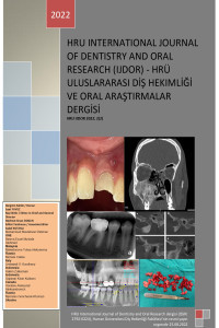
Panoramic radiograph (PR) is widely used in dentistry, yet certain anatomical variations can mimic pathological lesions. The aim of this case report is to present a maxillary sinus variation that appeared as a cyst-like lesion on PR around a previously endodontically treated tooth, and to highlight the diagnostic value of advanced imaging in such situations. A 31-year-old systemically healthy female patient presented with pain in the left posterior mandible. During routine PR examination, a well-defined radiolucent area was incidentally detected at the apex of the previously root canal–treated maxillary right second molar. Suspecting a periapical cyst, CBCT was performed and revealed no pathology related to the tooth; instead, multiple septa were identified within the maxillary sinus. One of these septa extended toward the tooth apex, confirming that the cyst-like appearance on PR resulted from anatomical variation combined with radiographic superimposition. This case illustrates that cyst-like areas observed on PRs are not always indicative of disease. Maxillary sinus septa and other anatomical variations can lead to diagnostic misinterpretation. Three-dimensional imaging plays a crucial role in differential diagnosis, helping clinicians avoid unnecessary invasive procedures.
Keywords
- Maxillary sinus
- anatomical variation
- periapical lesion
- cone-beam computed tomography
- panoramic radiography
Ethical Statement
References
- Assari A, Alotaibi N, Alajaji MA, Alqarni A, Alarishi MA. Characteristics of maxillary sinus septa: a cone-beam computed tomography evaluation. Int J Dent 2022;2022:2050257.
- Souza CFD, Loures ADO, Lopes DGF, Devito KL. Analysis of maxillary sinus septa by cone-beam computed tomography. Rev Odontol UNESP 2019;48:e20190034.
- Sekerci AE, Sisman Y, Etoz M, Bulut DG. Aberrant anatomical variation of maxillary sinus mimicking periapical cyst: a report of two cases and role of CBCT in diagnosis. Case Rep Dent 2013;2013:757645.
- Poleti ML, Fernandes LMPSR, Oliveira-Santos C, Capelozza AL, Chinellato LE, Rubira Bullen IR. Anatomical variation of the maxillary sinus in cone beam computed tomography. Case Rep Dent 2014;2014:707261.
- de Mesquita RA, Brasileiro CB, Amaral TMP. The importance of three-dimensional imaging from CBCT in elucidating a well-defined radiolucency image within the maxillary sinus: a case report. Open J Radiol 2014;4(3):255-9.
- Ricucci D, Pascon EA, Ford TR, Langeland K. Epithelium and bacteria in periapical lesions. Oral Surg Oral Med Oral Pathol Oral Radiol Endod 2006;101(2):239-49.
- Düker J. Radiographic diagnostics. Radicular cyst. Quintessence Int 2005;36(4):317.
- García CC, Sempere FV, Diago MP, Bowen EM. The post-endodontic periapical lesion: histologic and etiopathogenic aspects. Med Oral Pathol Oral Cir Bucal 2007;12:585-90.
Details
Primary Language
English
Subjects
Oral and Maxillofacial Radiology, Oral Medicine and Pathology
Journal Section
Case Report
Authors
Gözde Serindere
0000-0001-7439-3554
Türkiye
Publication Date
August 31, 2026
Submission Date
December 6, 2025
Acceptance Date
February 27, 2026
Published in Issue
Year 2026 Volume: 6 Number: 2