EN
Sentinel Lymph Node Mapping with Radiocolloid in Malign Melanoma: Significance of Histopathological Findings in Follow up
Abstract
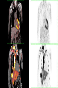
The presence of metastasis in regional lymph nodes has an essential value on survival in patients with malign melanoma. This study aimed to evaluate our experience on sentinel lymph node (SLN) mapping with radiocolloid in malign melanoma patients using SPECT/CT and present follow-up findings. Conventional planar lymphoscintigraphy and SPECT/CT images of 66 patients with primary cutaneous melanoma who underwent SLN biopsy in addition to the local primary excision or reexcision were retrospectively reviewed. All detected lymph nodes were removed and evaluated histopathologically. The number and histopathological findings of SLNs were determined, and follow-up findings of patients with metastatic or nonmetastatic nodes were comparedA statistically significant difference was not found in the number of detected SLN between planar and SPECT/CT imaging. The difference of total numbers of the metastatic and benign lymph nodes was statistically significant. A total of 55/66 patients were followed up with 10 months of median follow-up time and the presence of metastatic SLN on initial histopathological assessment was found to have a high risk for further metastatic disease. In the follow-up findings of 8/10 patients in whom SLN could not be detected on both planar and SPECT/CT imaging could be reached, normal findings were detected in 4 patients, and common nodal-visceral metastases in 4 patients. We concluded that, patients with malignant SLN histopathology had a high risk of developing metastases and should be followed up more frequently. As determined high metastases rate in patients with non-visualized SLN on radiocolloid imaging, it is essential to follow up also these patients meticulously.
Keywords
References
- Alazraki, N. P., Eshima, D., Eshima, L. A., Herda, S. C., Murray, D. R., Vansant, J. P., & Taylor, A. T. (1997). Lymphoscintigraphy, the sentinel node concept, and the intraoperative gamma probe in melanoma, breast cancer, and other potential cancers. Seminars in nuclear medicine, 27(1), 55–67. https://doi.org/10.1016/s0001-2998(97)80036-0
- Balch, C. M., Gershenwald, J. E., Soong, S. J., Thompson, J. F., Atkins, M. B., Byrd, D. R., Buzaid, A. C., Cochran, A. J., Coit, D. G., Ding, S., Eggermont, A. M., Flaherty, K. T., Gimotty, P. A., Kirkwood, J. M., McMasters, K. M., Mihm, M. C., Jr, Morton, D. L., Ross, M. I., Sober, A. J., & Sondak, V. K. (2009). Final version of 2009 AJCC melanoma staging and classification. Journal of clinical oncology : official journal of the American Society of Clinical Oncology, 27(36), 6199–6206. https://doi.org/10.1200/JCO.2009.23.4799
- Balch, C. M., Gershenwald, J. E., Soong, S. J., Thompson, J. F., Ding, S., Byrd, D. R., Cascinelli, N., Cochran, A. J., Coit, D. G., Eggermont, A. M., Johnson, T., Kirkwood, J. M., Leong, S. P., McMasters, K. M., Mihm, M. C., Jr, Morton, D. L., Ross, M. I., & Sondak, V. K. (2010). Multivariate analysis of prognostic factors among 2,313 patients with stage III melanoma: comparison of nodal micrometastases versus macrometastases. Journal of clinical oncology : official journal of the American Society of Clinical Oncology, 28(14), 2452–2459. https://doi.org/10.1200/JCO.2009.27.1627
- Benke, M., Wocial, K., Lewandowska, W., Rutkowski, P., Teterycz, P., Jarek, P., & Dedecjus, M. (2018). Value of planar lymphoscintigraphy (PL) versus SPECT/CT in evaluation of sentinel lymph node in trunk melanoma - one center, large series retrospective study. Nuclear medicine review. Central & Eastern Europe, 21(2), 79–84. https://doi.org/10.5603/NMR.a2018.0022
- Boland, G. M., & Gershenwald, J. E. (2012). Sentinel lymph node biopsy in melanoma. Cancer journal (Sudbury, Mass.), 18(2), 185–191. https://doi.org/10.1097/PPO.0b013e31825046c7
- Doepker, M. P., Yamamoto, M., Applebaum, M. A., Patel, N. U., Jaime Montilla-Soler, M., Sarnaik, A. A., Wayne Cruse, C., Sondak, V. K., & Zager, J. S. (2017). Comparison of Single-Photon Emission Computed Tomography-Computed Tomography (SPECT/CT) and Conventional Planar Lymphoscintigraphy for Sentinel Node Localization in Patients with Cutaneous Malignancies. Annals of surgical oncology, 24(2), 355–361. https://doi.org/10.1245/s10434-016-5590-8
- Erman, A. B., Collar, R. M., Griffith, K. A., Lowe, L., Sabel, M. S., Bichakjian, C. K., Wong, S. L., McLean, S. A., Rees, R. S., Johnson, T. M., & Bradford, C. R. (2012). Sentinel lymph node biopsy is accurate and prognostic in head and neck melanoma. Cancer, 118(4), 1040–1047. https://doi.org/10.1002/cncr.26288
- Gonzalez A. (2018). Sentinel Lymph Node Biopsy: Past and Present Implications for the Management of Cutaneous Melanoma with Nodal Metastasis. American journal of clinical dermatology, 19(Suppl 1), 24–30. https://doi.org/10.1007/s40257-018-0379-0
Details
Primary Language
English
Subjects
Nuclear Medicine
Journal Section
Research Article
Publication Date
September 6, 2024
Submission Date
August 7, 2024
Acceptance Date
August 20, 2024
Published in Issue
Year 2024 Volume: 4 Number: 2
APA
Uçak Semirgin, S., & Kirtiloglu, B. (2024). Sentinel Lymph Node Mapping with Radiocolloid in Malign Melanoma: Significance of Histopathological Findings in Follow up. Molecular Oncologic Imaging, 4(2), 1-9. https://izlik.org/JA38HB82YA
AMA
1.Uçak Semirgin S, Kirtiloglu B. Sentinel Lymph Node Mapping with Radiocolloid in Malign Melanoma: Significance of Histopathological Findings in Follow up. Molecular Oncologic Imaging. 2024;4(2):1-9. https://izlik.org/JA38HB82YA
Chicago
Uçak Semirgin, Sibel, and Banu Kirtiloglu. 2024. “Sentinel Lymph Node Mapping With Radiocolloid in Malign Melanoma: Significance of Histopathological Findings in Follow up”. Molecular Oncologic Imaging 4 (2): 1-9. https://izlik.org/JA38HB82YA.
EndNote
Uçak Semirgin S, Kirtiloglu B (September 1, 2024) Sentinel Lymph Node Mapping with Radiocolloid in Malign Melanoma: Significance of Histopathological Findings in Follow up. Molecular Oncologic Imaging 4 2 1–9.
IEEE
[1]S. Uçak Semirgin and B. Kirtiloglu, “Sentinel Lymph Node Mapping with Radiocolloid in Malign Melanoma: Significance of Histopathological Findings in Follow up”, Molecular Oncologic Imaging, vol. 4, no. 2, pp. 1–9, Sept. 2024, [Online]. Available: https://izlik.org/JA38HB82YA
ISNAD
Uçak Semirgin, Sibel - Kirtiloglu, Banu. “Sentinel Lymph Node Mapping With Radiocolloid in Malign Melanoma: Significance of Histopathological Findings in Follow up”. Molecular Oncologic Imaging 4/2 (September 1, 2024): 1-9. https://izlik.org/JA38HB82YA.
JAMA
1.Uçak Semirgin S, Kirtiloglu B. Sentinel Lymph Node Mapping with Radiocolloid in Malign Melanoma: Significance of Histopathological Findings in Follow up. Molecular Oncologic Imaging. 2024;4:1–9.
MLA
Uçak Semirgin, Sibel, and Banu Kirtiloglu. “Sentinel Lymph Node Mapping With Radiocolloid in Malign Melanoma: Significance of Histopathological Findings in Follow up”. Molecular Oncologic Imaging, vol. 4, no. 2, Sept. 2024, pp. 1-9, https://izlik.org/JA38HB82YA.
Vancouver
1.Sibel Uçak Semirgin, Banu Kirtiloglu. Sentinel Lymph Node Mapping with Radiocolloid in Malign Melanoma: Significance of Histopathological Findings in Follow up. Molecular Oncologic Imaging [Internet]. 2024 Sep. 1;4(2):1-9. Available from: https://izlik.org/JA38HB82YA