Research Article

PET/CT Imaging in Cardiac Sarcoidosis

Volume: 4 Number: 3 January 14, 2025
EN

PET/CT Imaging in Cardiac Sarcoidosis

Abstract

Sarcoidosis is a systemic granulomatous disease of unknown cause. The most commonly affected structures are the thoracic lymph nodes and the lungs, but any organ can be involved . In sarcoidosis, the heart can be affected both due to lung involvement and direct disease involvement. Clinically detected cardiac involvement is around 5%; However higher rates are reported in autopsy series. Endomyocardial biopsy, which is the definitive diagnostic method, is difficult and has low diagnostic value. Effective and reliable advanced cardiac imaging methods are needed in cardiac sarcoidosis . As well as the diagnosis of cardiac sarcoidosis, it is also important to determine disease activation, determine the treatment strategy, evaluate the severity, estimate the prognosis, and adjust steroid therapy . The most commonly used molecular imaging method in clinical practice for these purposes is Fluorodeoxyglucose (FDG) PET/CT imaging labeled with Fluorine-18 (F-18). Increased uptake in the myocardium on F-18 FDG PET/CT imaging is considered an important finding reflecting the activity of inflammation in cardiac sarcoidosis. In this report, two patients whose sarcoidosis diagnosis was confirmed histopathologically and who underwent PET CT examination due to cardiac sarcoidosis in our department are presented.

Keywords

References

  1. Handa, T., Nagai, S., Miki, S., Fushimi, Y., Ohta, K., Mishima, M., & Izumi, T. (2006). Incidence of pulmonary hypertension and its clinical relevance in patients with sarcoidosis. Chest, 129(5), 1246–1252. https://doi.org/10.1378/chest.129.5.1246
  2. Musellim, B., Kumbasar, O. O., Ongen, G., Cetinkaya, E., Turker, H., Uzaslan, E., Yenturk, E., Uzun, O., Saglam, L., Celik, G., Okumus, G., Annakkaya, A. N., Altiay, G., Tabak, L., Sakar, A., Kiter, G., Erturan, S., Turktas, H., Yalniz, E., Akkoclu, A., … Uzel, I. (2009). Epidemiological features of Turkish patients with sarcoidosis. Respiratory medicine, 103(6), 907–912. https://doi.org/10.1016/j.rmed.2008.12.011
  3. Fahy, G. J., Marwick, T., McCreery, C. J., Quigley, P. J., & Maurer, B. J. (1996). Doppler echocardiographic detection of left ventricular diastolic dysfunction in patients with pulmonary sarcoidosis. Chest, 109(1), 62–66. https://doi.org/10.1378/chest.109.1.62
  4. Mikami, R., Sekiguchi, M., Ryuzin, Y., Kobayashi, F., Hiraga, Y., Shimada, Y., Mochizuki, I., Kobayashi, T., Tamura, S., & Hosoda, Y. (1986). Changes in the peripheral vasculature of various organs in patients with sarcoidosis--possible role of microangiopathy. Heart and vessels, 2(3), 129–139. https://doi.org/10.1007/BF02128138
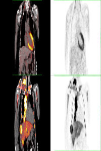
  5. Koiwa, H., Tsujino, I., Ohira, H., Yoshinaga, K., Otsuka, N., & Nishimura, M. (2010). Images in cardiovascular medicine: Imaging of cardiac sarcoid lesions using fasting cardiac 18F-fluorodeoxyglucose positron emission tomography: an autopsy case. Circulation, 122(5), 535–536. https://doi.org/10.1161/CIRCULATIONAHA.110.952184.
  6. Grutter JC, Drent M, van den Bosch JMM. Sarcoidosis. Eur Respir Mon 2009;46: 126-154.
  7. Yazaki, Y., Isobe, M., Hiroe, M., Morimoto, S., Hiramitsu, S., Nakano, T., Izumi, T., Sekiguchi, M., & Central Japan Heart Study Group (2001). Prognostic determinants of long-term survival in Japanese patients with cardiac sarcoidosis treated with prednisone. The American journal of cardiology, 88(9), 1006–1010. https://doi.org/10.1016/s0002-9149(01)01978-6
  8. Fleming, H. A., & Bailey, S. M. (1986). The prognosis of sarcoid heart disease in the United Kingdom. Annals of the New York Academy of Sciences, 465, 543–550. https://doi.org/10.1111/j.1749-6632.1986.tb18531.x

Details

Primary Language

English

Subjects

Nuclear Medicine

Journal Section

Research Article

Publication Date

January 14, 2025

Submission Date

December 25, 2024

Acceptance Date

December 27, 2024

Published in Issue

Year 2024 Volume: 4 Number: 3

APA
Gümüş, A., Özcan, P. P., Koç, Z. P., Süleymanoğlu, M., & Sağlam, Z. S. (2025). PET/CT Imaging in Cardiac Sarcoidosis. Molecular Oncologic Imaging, 4(3), 46-52. https://doi.org/10.71286/moi.1607134
AMA
1.Gümüş A, Özcan PP, Koç ZP, Süleymanoğlu M, Sağlam ZS. PET/CT Imaging in Cardiac Sarcoidosis. Molecular Oncologic Imaging. 2025;4(3):46-52. doi:10.71286/moi.1607134
Chicago
Gümüş, Adil, Pınar Pelin Özcan, Zehra Pınar Koç, Mahmut Süleymanoğlu, and Zeynep Selcan Sağlam. 2025. “PET/CT/Imaging/in/Cardiac/Sarcoidosis”. Molecular Oncologic Imaging 4 (3): 46-52. https://doi.org/10.71286/moi.1607134.
EndNote
Gümüş A, Özcan PP, Koç ZP, Süleymanoğlu M, Sağlam ZS (January 1, 2025) PET/CT Imaging in Cardiac Sarcoidosis. Molecular Oncologic Imaging 4 3 46–52.
IEEE
[1]A. Gümüş, P. P. Özcan, Z. P. Koç, M. Süleymanoğlu, and Z. S. Sağlam, “PET/CT Imaging in Cardiac Sarcoidosis”, Molecular Oncologic Imaging, vol. 4, no. 3, pp. 46–52, Jan. 2025, doi: 10.71286/moi.1607134.
ISNAD
Gümüş, Adil - Özcan, Pınar Pelin - Koç, Zehra Pınar - Süleymanoğlu, Mahmut - Sağlam, Zeynep Selcan. “PET/CT/Imaging/in/Cardiac/Sarcoidosis”. Molecular Oncologic Imaging 4/3 (January 1, 2025): 46-52. https://doi.org/10.71286/moi.1607134.
JAMA
1.Gümüş A, Özcan PP, Koç ZP, Süleymanoğlu M, Sağlam ZS. PET/CT Imaging in Cardiac Sarcoidosis. Molecular Oncologic Imaging. 2025;4:46–52.
MLA
Gümüş, Adil, et al. “PET/CT/Imaging/in/Cardiac/Sarcoidosis”. Molecular Oncologic Imaging, vol. 4, no. 3, Jan. 2025, pp. 46-52, doi:10.71286/moi.1607134.
Vancouver
1.Adil Gümüş, Pınar Pelin Özcan, Zehra Pınar Koç, Mahmut Süleymanoğlu, Zeynep Selcan Sağlam. PET/CT Imaging in Cardiac Sarcoidosis. Molecular Oncologic Imaging. 2025 Jan. 1;4(3):46-52. doi:10.71286/moi.1607134