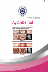
TREATMENT AND FOLLOW-UP OF EXTRUDED MAXILLARY CENTRAL INCISOR TEETH AS A RESULT OF TRAUMATIC DENTAL INJURY: CASE REPORT
Abstract
Extrusive luxation injury is a tooth injury
characterized by partial or complete separation
of the periodontal ligament (PDL), which causes
the tooth to displace in the axial direction. In
this article, the treatment of extruded maxillary
central incisor teeth as a result of trauma and twoyear
follow-up results are presented. In the case,
a 9-year-old female patient fell to the concrete
floor while walking on the road and there was an
extrusive luxation injury in the maxillary central
incisor teeth. The traumatized tooth was stabilized
with adjacent teeth by splinting and the patient was
followed up. After 2 weeks, 2 months, 6 months
and 2 year follow-up, the prognosis of the tooth
was examined clinically and radiographically.
This case report shows the importance of longterm
clinical and radiological follow-up in terms
of complications that may arise with the correct
treatment approach and follow-up program in
extrusive luxation injuries.
References
- [1] Bijella MFTB, Yared FNFG, Bijella VT, Lopes ES. Occurrence of primary incisor traumatism in Brazilian children: a houseby- house survey. ASDC J Dent Child 1990; 57:424–7. [2] Glendor U. Epidemiology of traumatic dental injuries - A 12 year review of the literature. Dent Traumatol 2008; 24:603‐11. [3] Hermann NV, Lauridsen E, Ahrensburg SS, Gerds TA, Andreasen JO. Periodontal healing complications following extrusive and lateral luxation in the permanent dentition: A longitudinal cohort study. Dent Traumatol 2012; 28:394‐402. [4] Andreasen FM, Andreasen JO. Textbook and color atlas of traumatic injuries to the teeth. 4 th ed. Oxford: Blackwell, 2007: 411–27. [5] Rock WP, Grundy MC. The effect of luxation and subluxation upon the prognosis of traumatized incisor teeth. J Dent 1981; 9:224‐30. [6] Schindler WG, Gullickson DC. Rationale for the management of calcific metamorphosis secondary to traumatic injuries. J Endod 1988; 14(8):408-12. [7] Amir FA, Gutmann JL, Witherspoon DE. Calcific metamorphosis: A challenge in endodontic diagnosis and treatment. Quintessence Int 2001; 32:447‐55. [8] Munley P, Goodell G. Calcific metamorphosis. Clinical Update for Naval Postgraduate Dental School. 2005; 27(4). [9] Lauridsen EF, Hermann NV, Gerds TA, Ahrensburg SS, Andreasen JO. Crown fractures Part 4 – healing complications of the pulp in permanent incisors following crown fractures with concurrent extrusion or lateral luxation injury. Dent Traumatol 2011;27. [10] Andreasen JO, Andreasen FM, Skeie A, Hjorting-Hansen E, Schwartz O. Effect of treatment delay upon pulp and periodontal healing of traumatic dental injuries – a review article. Dent Traumatol 2012; 18:116-28. [11] Andreasen FM. Pulpal healing after luxation injuries and root fracture in the permanent dentition. Endod Dent Traumatol 1989; 5:111–31. [12] Lee R, Barrett EJ, Kenny DJ. Clinical outcomes for permanent incisor luxations in a pediatric population. II. Extrusions. Dental Trau- matology. 2003; 19:274-9. [13] Lin S, Pilosof N, Karawani M, Wigler R, Kaufman AY, Teich ST. Occurrence and timing of complications following traumatic dental injuries: A retrospective study in a dental trauma department. J Clin Exp Dent 2016; 8(4):429-36. [14] Andreasen FM, Yu Z, Thomsen ML, Andersen PK. Occurrence of pulp canal obliteration after luxation injuries in the permanent dentition. Endod Dent Traumatol 1987; 3:103-15. [15] Bastos JV, Cortes MIS. Pulp canal obliteration after traumatic injuries in permanent teeth – scientific fact or fiction? Braz Oral Res 2018; 32:159-68.
TRAVMATİK DİŞ YARALANMASI SONUCU EKSTRÜZE OLMUŞ ÜST SANTRAL KESİCİ DİŞİN TEDAVİ VE TAKİBİ: OLGU SUNUMU
Abstract
Ekstrüziv lüksasyon yaralanması, dişin aksiyal yönde gevşemesine ve yer değiştirmesine neden olan periodontal ligamentin (PDL), kısmen veya tamamen ayrılması ile karakterize bir diş yaralanmasıdır. Bu makalede, travma sonucu üst sol santral kesici dişte meydana gelen ekstrüziv lüksasyon yaralanmasının tedavisi ve iki yıllık takip sonuçları sunulmaktadır. Olguda, 9 yasındaki kadın hasta, yolda yürürken beton zemine düşmüş ve üst sol santral kesici dişte ekstrüriv lüksasyon yaralanması meydana gelmiştir. Travmaya uğramış dişin komşu dişlere splintle stabilizasyonu sağlanmış ve hasta takibe alınmıştır. 2 hafta, 2 ay, 6 ay ve 2 senelik takipler sonucunda dişin klinik ve radyografik olarak prognozu incelenmiştir. Bu olgu sunumu, ekstrüziv lüksasyon yaralanmalarından sonra doğru tedavi yaklaşımı ve takip programı ile ortaya çıkabilecek komplikasyonlar açısından uzun süreli klinik ve radyolojik takibin önemini göstermektedir.
References
- [1] Bijella MFTB, Yared FNFG, Bijella VT, Lopes ES. Occurrence of primary incisor traumatism in Brazilian children: a houseby- house survey. ASDC J Dent Child 1990; 57:424–7. [2] Glendor U. Epidemiology of traumatic dental injuries - A 12 year review of the literature. Dent Traumatol 2008; 24:603‐11. [3] Hermann NV, Lauridsen E, Ahrensburg SS, Gerds TA, Andreasen JO. Periodontal healing complications following extrusive and lateral luxation in the permanent dentition: A longitudinal cohort study. Dent Traumatol 2012; 28:394‐402. [4] Andreasen FM, Andreasen JO. Textbook and color atlas of traumatic injuries to the teeth. 4 th ed. Oxford: Blackwell, 2007: 411–27. [5] Rock WP, Grundy MC. The effect of luxation and subluxation upon the prognosis of traumatized incisor teeth. J Dent 1981; 9:224‐30. [6] Schindler WG, Gullickson DC. Rationale for the management of calcific metamorphosis secondary to traumatic injuries. J Endod 1988; 14(8):408-12. [7] Amir FA, Gutmann JL, Witherspoon DE. Calcific metamorphosis: A challenge in endodontic diagnosis and treatment. Quintessence Int 2001; 32:447‐55. [8] Munley P, Goodell G. Calcific metamorphosis. Clinical Update for Naval Postgraduate Dental School. 2005; 27(4). [9] Lauridsen EF, Hermann NV, Gerds TA, Ahrensburg SS, Andreasen JO. Crown fractures Part 4 – healing complications of the pulp in permanent incisors following crown fractures with concurrent extrusion or lateral luxation injury. Dent Traumatol 2011;27. [10] Andreasen JO, Andreasen FM, Skeie A, Hjorting-Hansen E, Schwartz O. Effect of treatment delay upon pulp and periodontal healing of traumatic dental injuries – a review article. Dent Traumatol 2012; 18:116-28. [11] Andreasen FM. Pulpal healing after luxation injuries and root fracture in the permanent dentition. Endod Dent Traumatol 1989; 5:111–31. [12] Lee R, Barrett EJ, Kenny DJ. Clinical outcomes for permanent incisor luxations in a pediatric population. II. Extrusions. Dental Trau- matology. 2003; 19:274-9. [13] Lin S, Pilosof N, Karawani M, Wigler R, Kaufman AY, Teich ST. Occurrence and timing of complications following traumatic dental injuries: A retrospective study in a dental trauma department. J Clin Exp Dent 2016; 8(4):429-36. [14] Andreasen FM, Yu Z, Thomsen ML, Andersen PK. Occurrence of pulp canal obliteration after luxation injuries in the permanent dentition. Endod Dent Traumatol 1987; 3:103-15. [15] Bastos JV, Cortes MIS. Pulp canal obliteration after traumatic injuries in permanent teeth – scientific fact or fiction? Braz Oral Res 2018; 32:159-68.
Details
| Primary Language | Turkish |
|---|---|
| Subjects | Health Care Administration |
| Journal Section | Case Report |
| Authors | |
| Publication Date | October 1, 2019 |
| Submission Date | July 5, 2019 |
| Published in Issue | Year 2019 Volume: 5 Issue: 2 |
All site content, except where otherwise noted, is licensed under a Creative Common Attribution Licence. (CC-BY-NC 4.0)

