Abstract
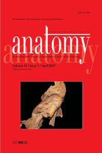
An understanding of the variations in the blood supply of the foregut and midgut are of critical importance to surgeons performing transplants, liver and biliary surgery, resection of tumors and various gastrointestinal procedures, as well as to interventional radiologists engaged in vessel embolization. During the dissection of a 95-year-old female cadaver as part of a course in medical gross anatomy at the University of California at Davis a rare series of vascular variations were observed. The left gastric artery arose independently from the abdominal aorta at the location of a typical celiac trunk. The common hepatic artery and splenic artery branched from a common vessel originating from a hepatosplenomesenteric trunk. Just inferior to the hepatosplenic trunk a hepatocolic trunk, which gave rise to an accessory right hepatic artery, dorsal pancreatic artery and a wandering mesenteric artery, branched from the superior mesenteric artery. This rare combination of clinically relevant variations was likely due to the abnormal partitioning and regression of the primitive splanchnic arteries during embryonic development.
Keywords
anatomical variation arc of Riolan celiac trunk celiacomesenteric trunk superior mesenteric artery
References
- 1. Haller AV. Icones anatomicae in quibus aliquae partes corporis humani delineatae proponuntur et arteriarum potissimum historia continetur. Göttingen: A. Vandenhoeck; 1756.
- 2. Chen H, Yano R, Emura S, Shoumura S. Anatomic variation of the celiac trunk with special reference to hepatic artery patterns. Ann Anat 2009;191:399–407.
- 3. Michels NA, Siddharth P, Kornblith PL, Parke WW. The variant blood supply to the descending colon, rectosigmoid and rectum based on 400 dissections. Its importance in regional resections: a review of medical literature. Dis Colon Rectum 1965;8:251-78.
- 4. Varotti G, Gondolesi GE, Goldman J, Wayne M, Florman SS, Schwartz ME, Miller CM, Sukru E. Anatomic variations in right liver living donors. J Am Coll Surg 2004;198:577–82.
- 5. Panagouli E, Venieratos D, Lolis E, Skandalakis P. Variations in the anatomy of the celiac trunk: a systematic review and clinical implications. Ann Anat 2013;195:501–11.
- 6. Sekiya S, Horiguchi M, Komatsu H, Kowada S, Yokoyama S, Yoshida K, Isogai S, Nakano M, Koizumi M. Persistent primitive sciatic artery associated with other various anomalies of vessels. Acta Anat (Basel) 1997;158:143–9.
- 7. Sakakibara K, Shindo S, Matsumoto M, Yoshida Y, Kimura M, Honda Y, Kamiya K, Katsu M, Kaga S, Suzuki S. Splenic artery aneurysm of the hepatosplenomesenteric trunk. Ann Vasc Dis 2013; 6:730–3.
- 8. Maldjian PD, Chorney MA. Celiomesenteric and hepatosplenomesenteric trunks: characterization of two rare vascular anomalies with CT. Abdom Imaging 2015;40:1800–7.
- 9. Prasanna LC, Alva R, Sneha GK, Bhat KM. Rare variations in the origin, branching pattern and course of the celiac trunk: report of two cases. Malays J Med Sci 2016;23:77–81.
- 10. Sahni DA, Jit IB, Gupta CN, Gupta DM, Harjeet E. Branches of the splenic artery and splenic arterial segments. Clin Anat 2003;16:371– 7.
- 11. Matsumura H. The significance of the morphology of the dorsal pancreatic artery in determining the presence of the accessory right hepatic artery passing behind the portal vein. Kaibogaku Zasshi 1998;73:517–27.
- 12. Tandler J. Über die varietäten der Arteria coeliaca und deren Entwicklung. Anat Hefte 1904;25:473–500.
- 13. Cavdar S, Sehirli U, Pekin B. Celiacomesenteric trunk. Clin Anat 1997;10:231–4.
Abstract
References
- 1. Haller AV. Icones anatomicae in quibus aliquae partes corporis humani delineatae proponuntur et arteriarum potissimum historia continetur. Göttingen: A. Vandenhoeck; 1756.
- 2. Chen H, Yano R, Emura S, Shoumura S. Anatomic variation of the celiac trunk with special reference to hepatic artery patterns. Ann Anat 2009;191:399–407.
- 3. Michels NA, Siddharth P, Kornblith PL, Parke WW. The variant blood supply to the descending colon, rectosigmoid and rectum based on 400 dissections. Its importance in regional resections: a review of medical literature. Dis Colon Rectum 1965;8:251-78.
- 4. Varotti G, Gondolesi GE, Goldman J, Wayne M, Florman SS, Schwartz ME, Miller CM, Sukru E. Anatomic variations in right liver living donors. J Am Coll Surg 2004;198:577–82.
- 5. Panagouli E, Venieratos D, Lolis E, Skandalakis P. Variations in the anatomy of the celiac trunk: a systematic review and clinical implications. Ann Anat 2013;195:501–11.
- 6. Sekiya S, Horiguchi M, Komatsu H, Kowada S, Yokoyama S, Yoshida K, Isogai S, Nakano M, Koizumi M. Persistent primitive sciatic artery associated with other various anomalies of vessels. Acta Anat (Basel) 1997;158:143–9.
- 7. Sakakibara K, Shindo S, Matsumoto M, Yoshida Y, Kimura M, Honda Y, Kamiya K, Katsu M, Kaga S, Suzuki S. Splenic artery aneurysm of the hepatosplenomesenteric trunk. Ann Vasc Dis 2013; 6:730–3.
- 8. Maldjian PD, Chorney MA. Celiomesenteric and hepatosplenomesenteric trunks: characterization of two rare vascular anomalies with CT. Abdom Imaging 2015;40:1800–7.
- 9. Prasanna LC, Alva R, Sneha GK, Bhat KM. Rare variations in the origin, branching pattern and course of the celiac trunk: report of two cases. Malays J Med Sci 2016;23:77–81.
- 10. Sahni DA, Jit IB, Gupta CN, Gupta DM, Harjeet E. Branches of the splenic artery and splenic arterial segments. Clin Anat 2003;16:371– 7.
- 11. Matsumura H. The significance of the morphology of the dorsal pancreatic artery in determining the presence of the accessory right hepatic artery passing behind the portal vein. Kaibogaku Zasshi 1998;73:517–27.
- 12. Tandler J. Über die varietäten der Arteria coeliaca und deren Entwicklung. Anat Hefte 1904;25:473–500.
- 13. Cavdar S, Sehirli U, Pekin B. Celiacomesenteric trunk. Clin Anat 1997;10:231–4.
Details
| Primary Language | English |
|---|---|
| Subjects | Health Care Administration |
| Journal Section | Case Reports |
| Authors | |
| Publication Date | April 30, 2017 |
| Published in Issue | Year 2017 Volume: 11 Issue: 1 |
Cite
Anatomy is the official journal of Turkish Society of Anatomy and Clinical Anatomy (TSACA).


