Abstract
Supporting Institution
yok
References
- Klein J, Juratli TA, Weise M, Schackert G. A systematic review of the semi-sitting position in neurosurgical patients with patent foramen ovale: how frequent is paradoxical embolism? World Neurosurg 2018;115:196–200.
- Muth CM, Shank ES. Gas embolism. N Engl J Med 2000;342:476– 82.
- Bothma PA, Schlimp CJ. Retrograde cerebral venous gas embolism: are we missing too many cases? Br J Anaesth 2014;112:401–4.
- Allioui S, Zaimi S, Sninate S, Abdellaoui M. Air bubbles in the brain: retrograde venous gas embolism in the cavernous sinus. Radiol Case Rep 2020;15:1011–3.
- Chuang DY, Sundararajan S, Sundararajan VA, Feldman DI, Xiong W. Accidental air embolism. Stroke 2019;50:e183– e186.
- Ploner F, Saltuari L, Marosi MJ, Dolif R, Salsa A. Cerebral air emboli with use of central venous catheter in mobile patient. Lancet 1991;338:1331.
- Tran P, Reed EJM, Hahn F, Lambrecht JE, McClay JC, Omojola MF. Incidence, radiographical features, and proposed mechanism for pneumocephalus from intravenous injection of air. West J Emerg Med 2010;11:180–5.
- McMinn RMH (editor). Last’s anatomy. Revised 9th ed. Chatswood: Elsevier Australia; 2019. p. 741–75.
- Standring S (editor). Gray’s anatomy: the anatomical basis of clinical practice. 40th ed. London: Churchill Livingstone Elsevier; 2008. p. 584.
- Souza MP, Magalhães E, Cascudo Ede F, Jogaib MA, Silva MC. Accidental catheterization of epidural venous plexus: tomographic analysis. Rev Bras Anestesiol 2016;66:208–11.
- Lanzieri CF. The significance of asymmetry of the foramen of Vesalius. AJNR Am J Neuroradiol 1998;9:1201–4.
Abstract
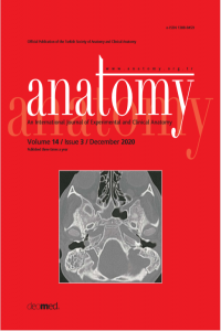
Pneumocephalus due to cerebral venous air embolism is an uncommon phenomenon. It results from retrograde progression of low weight air bubbles into dural venous sinuses during manipulation of a venous catheter, more frequently a central venous catheter through the subclavian and the jugular veins. However, it may also occur in relation with a peripheral intravenous catheter as in our case. We report a 91 year old female patient with congestive heart failure who had been examined in our emergency department two days previously due to dyspnea and received diuretic treatment through a peripheral intravenous line. She presented with vomiting and headache without obvious neurological deficits. Non-contrast cranial CT scan revealed wide spread punctate air bubbles inside and outside the cranial vault (pneumocephalus), within the venous system. The pneumocephalus was considered as iatrogenic due to the previous peripheral venous catheterization that resulted in retrograde migration of air bubbles through various venous connections into dural venous sinuses and extracranial veins. Since cerebral venous air embolism is a potentially serious complication of various medical procedures, it should be considered in differential diagnosis of nontraumatic headache and vomiting especially when there is a recent manipulation of venous lines. Cranial CT scan is helpful for early diagnosis.
References
- Klein J, Juratli TA, Weise M, Schackert G. A systematic review of the semi-sitting position in neurosurgical patients with patent foramen ovale: how frequent is paradoxical embolism? World Neurosurg 2018;115:196–200.
- Muth CM, Shank ES. Gas embolism. N Engl J Med 2000;342:476– 82.
- Bothma PA, Schlimp CJ. Retrograde cerebral venous gas embolism: are we missing too many cases? Br J Anaesth 2014;112:401–4.
- Allioui S, Zaimi S, Sninate S, Abdellaoui M. Air bubbles in the brain: retrograde venous gas embolism in the cavernous sinus. Radiol Case Rep 2020;15:1011–3.
- Chuang DY, Sundararajan S, Sundararajan VA, Feldman DI, Xiong W. Accidental air embolism. Stroke 2019;50:e183– e186.
- Ploner F, Saltuari L, Marosi MJ, Dolif R, Salsa A. Cerebral air emboli with use of central venous catheter in mobile patient. Lancet 1991;338:1331.
- Tran P, Reed EJM, Hahn F, Lambrecht JE, McClay JC, Omojola MF. Incidence, radiographical features, and proposed mechanism for pneumocephalus from intravenous injection of air. West J Emerg Med 2010;11:180–5.
- McMinn RMH (editor). Last’s anatomy. Revised 9th ed. Chatswood: Elsevier Australia; 2019. p. 741–75.
- Standring S (editor). Gray’s anatomy: the anatomical basis of clinical practice. 40th ed. London: Churchill Livingstone Elsevier; 2008. p. 584.
- Souza MP, Magalhães E, Cascudo Ede F, Jogaib MA, Silva MC. Accidental catheterization of epidural venous plexus: tomographic analysis. Rev Bras Anestesiol 2016;66:208–11.
- Lanzieri CF. The significance of asymmetry of the foramen of Vesalius. AJNR Am J Neuroradiol 1998;9:1201–4.
Details
| Primary Language | English |
|---|---|
| Subjects | Health Care Administration |
| Journal Section | Case Reports |
| Authors | |
| Publication Date | December 30, 2020 |
| Published in Issue | Year 2020 Volume: 14 Issue: 3 |
Cite
Anatomy is the official journal of Turkish Society of Anatomy and Clinical Anatomy (TSACA).


