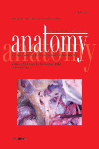
Clinical implications of the anatomical relationships of the pterygopalatine fossa and Vidian canal: an endonasal endoscopic cadaveric study
Abstract
Objectives: The aim of our study was to describe a surgical access to pterygopalatine fossa and Vidian canal using an endonasal endoscopic approach. We also intend to reveal the anatomical relations of the neurovascular structures in the surgical corridors and to determine the relationships between previously defined reference points in order to prevent surgical complications during surgical access to these regions.
Methods: Our study was carried out between October-December 2016 in Cerrahpaşa Faculty of Medicine Microneurosurgery and Neuroanatomy Laboratory. A total of 7 silicon dye-injected cadavers (4 males and 3 females) were studied. 3D images were obtained by photographing the approaches applied to the pterygopalatine fossa and Vidian canal and related anatomical structures.
Results: We succeeded in exposing and examining the pterygopalatine fossa and Vidian canal endoscopically in all samples. First, the posterior wall of the maxillary sinus was opened to reach the pterygopalatine fossa. The pterygopalatine fossa was divided into 3 anatomical compartments. The first layer encountered was the periosteum covering the pterygopalatine fossa. After removing the periosteum, the fat layer was revealed. Under the fat layer, the vascular compartment and finally the neural compartment were encountered.
Conclusion: Our study revealed three-dimensional anatomical data related to the surgical margins involved in approaches to the pterygopalatine fossa and Vidian canal; and specifically defined various neurovascular structures encountered in these approaches. Our study provides information to decrease potential complications that may develop during endonasal endoscopic surgery. We conclude that as the anatomy of the pterygopalatine fossa and Vidian canal is known in details, the endonasal endoscopic approach is likely to become the standard method to access lesions in these regions.
References
- Alfieri A, Jho HD, Schettino R, Tschabitscher M. Endoscopic endonasal approach to the pterygopalatine fossa: anatomic study. Neurosurgery 2003;52:374–80.
- Bahşi İ, Orhan M, Kervancıoğlu P, Yalçın ED. The anatomical and radiological evaluation of the Vidian canal on cone-beam computed tomography images. Eur Arch Otorhinolaryngol 2019;276:1373–83.
- Baldauf J, Hosemann W, Schroeder HW. Endoscopic endonasal approach for craniopharyngiomas. Neurosurg Clin N Am 2015;26: 363;75.
- Bolger WE. Endoscopic transpterygoid approach to the lateral sphenoid recess: surgical approach and clinical experience. Otolaryngol Head Neck Surg 2005;133:20–6.
- Buchmann L, Larsen C, Pollack A, Tawfik O, Sykes K, Hoover LA. Endoscopic techniques in resection of anterior skull base/paranasal sinus malignancies. Laryngoscope 2006;116:1749–54.
- Cappabianca P, Alfieri A, De Divitiis E. Endoscopic endonasal transsphenoidal approach to the sella: towards functional endoscopic pituitary surgery (FEPS). Minim Invasive Neursourg 1998;41:66–73.
- Cappabianca P, Cavallo LM, de Divitiis O, de Angelis M, Chiaramonte C, Solari D. Endoscopic endonasal extended approaches for the management of large pituitary adenomas. Neurosurg Clin N Am 2015;26:323–31.
- Choi J, Park HS. The clinical anatomy of the maxillary artery in the pterygopalatine fossa. J Oral Maxillofac Surg 2003;61:72–8.
- Dave SP, Bared A, Casiano RR. Surgical outcomes and safety of transnasal endoscopic resection for anterior skull tumors. J Otolaryngol Head Neck Surg 2007;136:920–7.
- De Divitiis E, Cappabianca P, Cavallo LM. Endoscopic transsphenoidal approach: adaptability of the procedure to different sellar lesions. Neurosurgery 2002;51:699–707.
- De Divitiis E, Cavallo LM, Cappabianca P, Esposito F. Extended endoscopic endonasal transsphenoidal approach for the removal of suprasellar tumors: part 2. Neurosurgery 2007;60:46–59.
- De Lara D, Ditzel Filho LF, Prevedello DM, Carrau RL, Kasemsiri P, Otto BA, Kassam AB. Endonasal endoscopic approaches to the paramedian skull base. World Neurosurg 2014;82:S121–9.
- Dehdashti AR, Ganna A, Witterick I, Gentili F. Expanded endoscopic endonasal approach for anterior cranial base and suprasellar lesions: indications and limitations. Neurosurgery 2009;64:677–89.
- Ditzel Filho LF, Prevedello DM, Dolci RL, Jamshidi AO, Kerr EE, Campbell R, Otto BA, Carrau RL. The endoscopic endonasal approach for removal of petroclival chondrosarcomas. Neurosurg Clin N Am 2015;26:453–62.
- Fortes FS, Sennes LU, Carrau RL, Brito R, Ribas GC, Yasuda A, Rodrigues Jr AJ, Synderman CH, Kassam AB. Endoscopic anatomy of the pterygopalatine fossa and the transpterygoid approach: development of a surgical instruction model. Laryngoscope 2008;118:44–9.
- Frank G, Pasquini E, Doglietto F, Mazzatenta D, Sciarretta V, Farneti G, Calbucci F. The endoscopic extended transsphenoidal approach for craniopharyngiomas. Neurosurgery 2006;ONS75–83.
- Har-El G. Combined endoscopic transmaxillary-transnasal approach to the pterygoid region, lateral sphenoid sinus, and retrobulbar orbit. Ann Otol Rhinol Laryngol 2005;114:439–42.
- Hofstetter CP, Singh A, Anand VK, Kacker A, Schwartz TH. The endoscopic, endonasal, transmaxillary transpterygoid approach to the pterygopalatine fossa, infratemporal fossa, petrous apex, and the Meckel cave. J Neurosurg 2010;113:967–74.
- Karkas A, Zimmer LA, Theodosopoulos PV, Keller JT, Prades JM. Endonasal endoscopic approach to the pterygopalatine and infratemporal fossae. Eur Ann Otorhinolaryngol Head Neck Dis 2021;138:391–5.
- Kassam AB, Gardner P, Snyderman C, Mintz A, Carrau R. Expanded endonasal approach: fully endoscopic, completely transnasal approach to the middle third of the clivus, petrous bone, middle cranial fossa, and infratemporal fossa. Neurosurg Focus 2005;19:E6.
- Kassam A B, Vescan AD, Carrau RL, Prevedello DM, Gardner P, Mintz AH, Synderman CH, Rhoton AL. Expanded endonasal approach: vidian canal as a landmark to the petrous internal carotid artery. J Neurosurg 2008;108:177–83.
- Klossek JM, Ferrie JC, Goujon JM, Fontanel JP. Endoscopic approach of the pterygopalatine fossa: report of one case. Rhinology 1994;32:208–10.
- Kutlay M, Durmaz A, Özer İ, Kural C, Temiz Ç, Kaya S, Solmaz İ, Daneyemez M, İzci Y. Extended endoscopic endonasal approach to the ventral skull base lesions. Clin Neurol Neurosurg 2018;167:129–40.
- Laufer I, Anand VK, Schwartz TH. Endoscopic, endonasal extended transsphenoidal, transplanum transtuberculum approach for resection of suprasellar lesions. J Neurosurg 2007;106:400–6.
- Lobo B, Zhang X, Barkhoudarian G, Griffiths CF, Kelly DF. Endonasal endoscopic management of parasellar and cavernous sinus meningiomas. Neurosurg Clin N Am 2015;26:389–401.
- Loukas M. Book review: clinically oriented anatomy, 5th ed. by Keith L. Moore and Arthur F. Dalley II. Clin Anat 2006;16:367
- Morton AL, Khan A. Internal maxillary artery variability in the pterygopalatine fossa. Otolaryngol Head Neck Surg 1991;104:204–9.
- Pinheiro-Neto CD, Fernandez-Miranda JC, Prevedello DM, Carrau RL, Gardner PA, Snyderman CH. Transposition of the pterygopalatine fossa during endoscopic endonasal transpterygoid approaches. J Neurol Surg B Skull Base 2013;74:266–70.
- Prevedello DM, Pinheiro-Neto CD, Fernandez-Miranda JC, Carrau RL, Snyderman CH, Gardner PA, Kassam AB. Vidian nerve transposition for endoscopic endonasal middle fossa approaches. Neurosurgery 2010;67:478–84.
- Schwartz TH, Fraser JF, Brown S, Tabaee A, Kacker A, Anand VK. Endoscopic cranial base surgery: classification of operative approaches. Neurosurgery 2008;62:991–1005.
- Snyderman C, Gardner P. Master techniques in otolaryngology-head and neck surgery: skull base surgery. Philadelphia (PA): Wolters Kluwer; 2015. p. 469.
- Vescan AD, Snyderman CH, Carrau RL, Mintz A, Gardner P, Branstetter 4th B, Kassam AB. Vidian canal: analysis and relationship to the internal carotid artery. Laryngoscope 2007;117:1338–42.
Abstract
References
- Alfieri A, Jho HD, Schettino R, Tschabitscher M. Endoscopic endonasal approach to the pterygopalatine fossa: anatomic study. Neurosurgery 2003;52:374–80.
- Bahşi İ, Orhan M, Kervancıoğlu P, Yalçın ED. The anatomical and radiological evaluation of the Vidian canal on cone-beam computed tomography images. Eur Arch Otorhinolaryngol 2019;276:1373–83.
- Baldauf J, Hosemann W, Schroeder HW. Endoscopic endonasal approach for craniopharyngiomas. Neurosurg Clin N Am 2015;26: 363;75.
- Bolger WE. Endoscopic transpterygoid approach to the lateral sphenoid recess: surgical approach and clinical experience. Otolaryngol Head Neck Surg 2005;133:20–6.
- Buchmann L, Larsen C, Pollack A, Tawfik O, Sykes K, Hoover LA. Endoscopic techniques in resection of anterior skull base/paranasal sinus malignancies. Laryngoscope 2006;116:1749–54.
- Cappabianca P, Alfieri A, De Divitiis E. Endoscopic endonasal transsphenoidal approach to the sella: towards functional endoscopic pituitary surgery (FEPS). Minim Invasive Neursourg 1998;41:66–73.
- Cappabianca P, Cavallo LM, de Divitiis O, de Angelis M, Chiaramonte C, Solari D. Endoscopic endonasal extended approaches for the management of large pituitary adenomas. Neurosurg Clin N Am 2015;26:323–31.
- Choi J, Park HS. The clinical anatomy of the maxillary artery in the pterygopalatine fossa. J Oral Maxillofac Surg 2003;61:72–8.
- Dave SP, Bared A, Casiano RR. Surgical outcomes and safety of transnasal endoscopic resection for anterior skull tumors. J Otolaryngol Head Neck Surg 2007;136:920–7.
- De Divitiis E, Cappabianca P, Cavallo LM. Endoscopic transsphenoidal approach: adaptability of the procedure to different sellar lesions. Neurosurgery 2002;51:699–707.
- De Divitiis E, Cavallo LM, Cappabianca P, Esposito F. Extended endoscopic endonasal transsphenoidal approach for the removal of suprasellar tumors: part 2. Neurosurgery 2007;60:46–59.
- De Lara D, Ditzel Filho LF, Prevedello DM, Carrau RL, Kasemsiri P, Otto BA, Kassam AB. Endonasal endoscopic approaches to the paramedian skull base. World Neurosurg 2014;82:S121–9.
- Dehdashti AR, Ganna A, Witterick I, Gentili F. Expanded endoscopic endonasal approach for anterior cranial base and suprasellar lesions: indications and limitations. Neurosurgery 2009;64:677–89.
- Ditzel Filho LF, Prevedello DM, Dolci RL, Jamshidi AO, Kerr EE, Campbell R, Otto BA, Carrau RL. The endoscopic endonasal approach for removal of petroclival chondrosarcomas. Neurosurg Clin N Am 2015;26:453–62.
- Fortes FS, Sennes LU, Carrau RL, Brito R, Ribas GC, Yasuda A, Rodrigues Jr AJ, Synderman CH, Kassam AB. Endoscopic anatomy of the pterygopalatine fossa and the transpterygoid approach: development of a surgical instruction model. Laryngoscope 2008;118:44–9.
- Frank G, Pasquini E, Doglietto F, Mazzatenta D, Sciarretta V, Farneti G, Calbucci F. The endoscopic extended transsphenoidal approach for craniopharyngiomas. Neurosurgery 2006;ONS75–83.
- Har-El G. Combined endoscopic transmaxillary-transnasal approach to the pterygoid region, lateral sphenoid sinus, and retrobulbar orbit. Ann Otol Rhinol Laryngol 2005;114:439–42.
- Hofstetter CP, Singh A, Anand VK, Kacker A, Schwartz TH. The endoscopic, endonasal, transmaxillary transpterygoid approach to the pterygopalatine fossa, infratemporal fossa, petrous apex, and the Meckel cave. J Neurosurg 2010;113:967–74.
- Karkas A, Zimmer LA, Theodosopoulos PV, Keller JT, Prades JM. Endonasal endoscopic approach to the pterygopalatine and infratemporal fossae. Eur Ann Otorhinolaryngol Head Neck Dis 2021;138:391–5.
- Kassam AB, Gardner P, Snyderman C, Mintz A, Carrau R. Expanded endonasal approach: fully endoscopic, completely transnasal approach to the middle third of the clivus, petrous bone, middle cranial fossa, and infratemporal fossa. Neurosurg Focus 2005;19:E6.
- Kassam A B, Vescan AD, Carrau RL, Prevedello DM, Gardner P, Mintz AH, Synderman CH, Rhoton AL. Expanded endonasal approach: vidian canal as a landmark to the petrous internal carotid artery. J Neurosurg 2008;108:177–83.
- Klossek JM, Ferrie JC, Goujon JM, Fontanel JP. Endoscopic approach of the pterygopalatine fossa: report of one case. Rhinology 1994;32:208–10.
- Kutlay M, Durmaz A, Özer İ, Kural C, Temiz Ç, Kaya S, Solmaz İ, Daneyemez M, İzci Y. Extended endoscopic endonasal approach to the ventral skull base lesions. Clin Neurol Neurosurg 2018;167:129–40.
- Laufer I, Anand VK, Schwartz TH. Endoscopic, endonasal extended transsphenoidal, transplanum transtuberculum approach for resection of suprasellar lesions. J Neurosurg 2007;106:400–6.
- Lobo B, Zhang X, Barkhoudarian G, Griffiths CF, Kelly DF. Endonasal endoscopic management of parasellar and cavernous sinus meningiomas. Neurosurg Clin N Am 2015;26:389–401.
- Loukas M. Book review: clinically oriented anatomy, 5th ed. by Keith L. Moore and Arthur F. Dalley II. Clin Anat 2006;16:367
- Morton AL, Khan A. Internal maxillary artery variability in the pterygopalatine fossa. Otolaryngol Head Neck Surg 1991;104:204–9.
- Pinheiro-Neto CD, Fernandez-Miranda JC, Prevedello DM, Carrau RL, Gardner PA, Snyderman CH. Transposition of the pterygopalatine fossa during endoscopic endonasal transpterygoid approaches. J Neurol Surg B Skull Base 2013;74:266–70.
- Prevedello DM, Pinheiro-Neto CD, Fernandez-Miranda JC, Carrau RL, Snyderman CH, Gardner PA, Kassam AB. Vidian nerve transposition for endoscopic endonasal middle fossa approaches. Neurosurgery 2010;67:478–84.
- Schwartz TH, Fraser JF, Brown S, Tabaee A, Kacker A, Anand VK. Endoscopic cranial base surgery: classification of operative approaches. Neurosurgery 2008;62:991–1005.
- Snyderman C, Gardner P. Master techniques in otolaryngology-head and neck surgery: skull base surgery. Philadelphia (PA): Wolters Kluwer; 2015. p. 469.
- Vescan AD, Snyderman CH, Carrau RL, Mintz A, Gardner P, Branstetter 4th B, Kassam AB. Vidian canal: analysis and relationship to the internal carotid artery. Laryngoscope 2007;117:1338–42.
Details
| Primary Language | English |
|---|---|
| Subjects | Brain and Nerve Surgery (Neurosurgery) |
| Journal Section | Original Articles |
| Authors | |
| Publication Date | December 20, 2022 |
| Published in Issue | Year 2022 Volume: 16 Issue: 3 |
Cite
Anatomy is the official journal of Turkish Society of Anatomy and Clinical Anatomy (TSACA).

