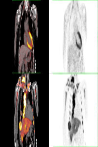
Abdominal Mass shown by the FDG PET/CT with diagnosis of Burkitt Lymphoma
Abstract
Keywords
References
- 1. Sachpekidis, C., Exadaktylou, P., Katsampoukas, D., Moralidis, E., & Arsos, G. (2020). 18F-FDG PET/CT in treatment response evaluation of Burkitt lymphoma: complete remission of a peritoneal super scan. Hellenic journal of nuclear medicine, 23(1), 76–78. https://doi.org/10.1967/s002449912006
- 2. Zeng, W., Lechowicz, M. J., Winton, E., Cho, S. M., Galt, J. R., & Halkar, R. (2009). Spectrum of FDG PET/CT findings in Burkitt lymphoma. Clinical nuclear medicine, 34(6), 355–358. https://doi.org/10.1097/RLU.0b013e3181a34552
- 3. Wang, X., Chen, Z., Tang, G., & Zhang, X. (2011). A child with Burkitt lymphoma with pleural, peritoneal, mesenteric, omental, and renal involvement: diagnostics by FDG PET/CT. Clinical nuclear medicine, 36(7), 612–615. https://doi.org/10.1097/RLU.0b013e318217af84
Details
Primary Language
English
Subjects
Oncology and Carcinogenesis
Journal Section
Case Report
Authors
Gökçe Yavan
*
This is me
0000-0002-7332-6293
Türkiye
Zehra Pınar Koç
This is me
0000-0002-3274-5790
Türkiye
Pınar Pelin Özcan
This is me
0000-0003-0147-2678
Türkiye
Publication Date
August 1, 2022
Submission Date
July 8, 2022
Acceptance Date
July 15, 2022
Published in Issue
Year 2022 Volume: 2 Number: 2