Research Article

Imaging Features of Chest Wall Mass in the Hybrid Imaging Modalities: SPECT/CT and PET/CT

Volume: 3 Number: 3 December 30, 2023
EN

Imaging Features of Chest Wall Mass in the Hybrid Imaging Modalities: SPECT/CT and PET/CT

Abstract

Background: The chest wall tumors are rare lesions thus there are limited literature data about the radiologic appearance of these tumors and no previous study exists about the diagnostic performance of the functional imaging modalities. Aim of this study is to analyze the diagnostic accuracy of the hybrid imaging modalities: Single photon emission computed tomography/computed tomography (SPECT/CT) and Positron emission tomography/computed tomography (PET/CT) in comparison to the histopathology results of the chest wall tumors. Materials and Methods: Imaging results of the patients (n=20, 7F/13M, mean: 53±21,9 years of age) with the diagnosis of chest wall mass were recorded. Retrospective evaluation of the images were performed by an experienced Nuclear Medicine physician and compared with the histopathology results as gold standard. Results: The lesions of the patients that were involved in this study were mean: 63±33.8 mm and the standardized uptake value (SUVmax) were mean: 13.7±10.8. The imaging/metabolic characteristics of the lesions were considered malign in all the patients included. All the patients diagnosed to have malign neoplasms except two. Conclusion: The hybrid imaging has excellent diagnostic facility in diagnosis of the chest wall neoplasm. Prospective studies about this issue are warranted.

Keywords

Ethical Statement

Ethics approval was obtained, consent to participate obtained, consent for publication obtained, availability of data and material provided, competing interests;nothing to disclose, funding; none provided, authors' contributions; sufficently participated, acknowledgements; none.

References

  1. Ilgan, S., Dikmen, E., Cetinkanat, C. G., Dakak, M., & Güngör, A. (2014). Schwannomatozis of the Chest Wall: FDG PET Findings. Molecular imaging and radionuclide therapy, 23(2), 64–66. https://doi.org/10.4274/mirt.353
  2. Chadaz, T., Hobbs, S. K., & Son, H. (2013). Chest wall sarcoma: 18F-FDG PET/CT in a patient with Li-Fraumeni syndrome. Clinical nuclear medicine, 38(10), 818–820. https://doi.org/10.1097/RLU.0b013e3182a20033
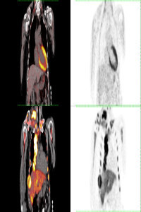
  3. Carter, B. W., Benveniste, M. F., Betancourt, S. L., de Groot, P. M., Lichtenberger, J. P., 3rd, Amini, B., & Abbott, G. F. (2016). Imaging Evaluation of Malignant Chest Wall Neoplasms. Radiographics : a review Molecular Oncologic Imaging 2023;3(3):01-07 7 publication of the Radiological Society of North America, Inc, 36(5), 1285–1306. https://doi.org/10.1148/rg.2016150208
  4. Jiang, L., Gao, Y., Sheng, S., Xu, M., Lu, L., & Lu, H. (2011). A first described chest wall metastasis from colon cancer demonstrated with (18)F-FDG PET/CT. Hellenic journal of nuclear medicine, 14(3), 316–317.
  5. Park BJ, Flores RM. Chest wall tumors. In: Shields TW, Locicero J, Reed CE, Feins RH, eds. General thoracic surgery. Philadelphia, Pa: Lippincott, 2009; 669–678.
  6. David, E. A., & Marshall, M. B. (2011). Review of chest wall tumors: a diagnostic, therapeutic, and reconstructive challenge. Seminars in plastic surgery, 25(1), 16–24. https://doi.org/10.1055/s-0031-1275167
  7. Mullan, C. P., Madan, R., Trotman-Dickenson, B., Qian, X., Jacobson, F. L., & Hunsaker, A. (2011). Radiology of chest wall masses. AJR. American journal of roentgenology, 197(3), W460–W470. https://doi.org/10.2214/AJR.10.7259
  8. Bandi, V., Lunn, W., Ernst, A., Eberhardt, R., Hoffmann, H., & Herth, F. J. (2008). Ultrasound vs. CT in detecting chest wall invasion by tumor: a prospective study. Chest, 133(4), 881–886. https://doi.org/10.1378/chest.07-1656

Details

Primary Language

English

Subjects

Thoracic Surgery, Nuclear Medicine

Journal Section

Research Article

Publication Date

December 30, 2023

Submission Date

November 15, 2023

Acceptance Date

December 25, 2023

Published in Issue

Year 2023 Volume: 3 Number: 3

APA
Koç, Z. P., Kara, P. Ö., Köksel, M. O., & Sayar, H. (2023). Imaging Features of Chest Wall Mass in the Hybrid Imaging Modalities: SPECT/CT and PET/CT. Molecular Oncologic Imaging, 3(3), 1-7. https://izlik.org/JA95CZ23KD
AMA
1.Koç ZP, Kara PÖ, Köksel MO, Sayar H. Imaging Features of Chest Wall Mass in the Hybrid Imaging Modalities: SPECT/CT and PET/CT. Molecular Oncologic Imaging. 2023;3(3):1-7. https://izlik.org/JA95CZ23KD
Chicago
Koç, Zehra Pınar, Pelin Özcan Kara, Mehmet Oğuz Köksel, and Hamide Sayar. 2023. “Imaging Features of Chest Wall Mass in the Hybrid Imaging Modalities: SPECT CT and PET CT”. Molecular Oncologic Imaging 3 (3): 1-7. https://izlik.org/JA95CZ23KD.
EndNote
Koç ZP, Kara PÖ, Köksel MO, Sayar H (December 1, 2023) Imaging Features of Chest Wall Mass in the Hybrid Imaging Modalities: SPECT/CT and PET/CT. Molecular Oncologic Imaging 3 3 1–7.
IEEE
[1]Z. P. Koç, P. Ö. Kara, M. O. Köksel, and H. Sayar, “Imaging Features of Chest Wall Mass in the Hybrid Imaging Modalities: SPECT/CT and PET/CT”, Molecular Oncologic Imaging, vol. 3, no. 3, pp. 1–7, Dec. 2023, [Online]. Available: https://izlik.org/JA95CZ23KD
ISNAD
Koç, Zehra Pınar - Kara, Pelin Özcan - Köksel, Mehmet Oğuz - Sayar, Hamide. “Imaging Features of Chest Wall Mass in the Hybrid Imaging Modalities: SPECT CT and PET CT”. Molecular Oncologic Imaging 3/3 (December 1, 2023): 1-7. https://izlik.org/JA95CZ23KD.
JAMA
1.Koç ZP, Kara PÖ, Köksel MO, Sayar H. Imaging Features of Chest Wall Mass in the Hybrid Imaging Modalities: SPECT/CT and PET/CT. Molecular Oncologic Imaging. 2023;3:1–7.
MLA
Koç, Zehra Pınar, et al. “Imaging Features of Chest Wall Mass in the Hybrid Imaging Modalities: SPECT CT and PET CT”. Molecular Oncologic Imaging, vol. 3, no. 3, Dec. 2023, pp. 1-7, https://izlik.org/JA95CZ23KD.
Vancouver
1.Zehra Pınar Koç, Pelin Özcan Kara, Mehmet Oğuz Köksel, Hamide Sayar. Imaging Features of Chest Wall Mass in the Hybrid Imaging Modalities: SPECT/CT and PET/CT. Molecular Oncologic Imaging [Internet]. 2023 Dec. 1;3(3):1-7. Available from: https://izlik.org/JA95CZ23KD