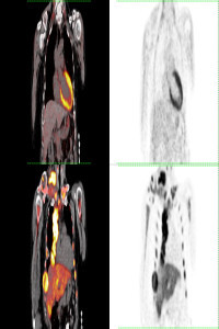
Vulva lesions as characterized by F-18 Fluorodeoxyglucose Positron Emission Tomography/Computed Tomography
Abstract
Keywords
References
- Brar, H., May, T., Tau, N., Langer, D., MacCrostie, P., Han, K., & Metser, U. (2017). Detection of extra-regional tumour recurrence with 18F-FDG-PET/CT in patients with recurrent gynaecological malignancies being considered for radical salvage surgery. Clinical radiology, 72(4), 302–306. https://doi.org/10.1016/j.crad.2016.12.009
- Rao, Y. J., Hassanzadeh, C., Chundury, A., Hui, C., Siegel, B. A., Dehdashti, F., DeWees, T., Mullen, D., Powell, M. A., Mutch, D. G., Schwarz, J. K., & Grigsby, P. W. (2017). Association of post-treatment positron emission tomography with locoregional control and survival after radiation therapy for squamous cell carcinoma of the vulva. Radiotherapy and oncology : journal of the European Society for Therapeutic Radiology and Oncology, 122(3), 445–451. https://doi.org/10.1016/j.radonc.2016.12.019
- Lin, G., Chen, C. Y., Liu, F. Y., Yang, L. Y., Huang, H. J., Huang, Y. T., Jung, S. M., Chou, H. H., Lai, C. H., & Ng, K. K. (2015). Computed tomography, magnetic resonance imaging and FDG positron emission tomography in the management of vulvar malignancies. European radiology, 25(5), 1267–1278. https://doi.org/10.1007/s00330-014-3530-1
- Shen, G., Wang, R., Pan, L., & Kuang, A. (2021). Malignant Extrarenal Rhabdoid Tumor of the Vagina on FDG PET/CT. Clinical nuclear medicine, 46(12), 1020–1021. https://doi.org/10.1097/RLU.0000000000003751
- Beyhan, E., Ergül, N., Bektaş, S., Erol, Ö., & Çermik, T. F. (2021). Solitary Vulvar Involvement of Ovarian Non-Hodgkin Lymphoma Mimicking Bartholin's Abscess on 18F-FDG PET/CT. Clinical nuclear medicine, 46(3), 255–257. https://doi.org/10.1097/RLU.0000000000003494
- Guglielmo, P., Paderno, M., Elisei, F., Guerra, L., Landoni, C., Buda, A., & Crivellaro, C. (2019). 18F-FDG PET/CT in a Case of Metastatic Breast Cancer to the Vulva. Clinical nuclear medicine, 44(7), 572–573. https://doi.org/10.1097/RLU.0000000000002583
- Gandhi, A. K., Roy, S., Mridha, A. R., & Sharma, D. N. (2015). Vulvar metastasis from carcinoma breast unveiling distant metastasis: Exploring an unusual metastatic pattern. Journal of the Egyptian National Cancer Institute, 27(4), 243–246. https://doi.org/10.1016/j.jnci.2015.05.005
- Treglia, G., Paone, G., Perriard, U., Ceriani, L., & Giovanella, L. (2014). An unusual case of diffuse large B-cell lymphoma involving the vulva evaluated by 18F-FDG PET/CT. Clinical nuclear medicine, 39(10), e439–e441. https://doi.org/10.1097/RLU.0000000000000258
Details
Primary Language
English
Subjects
Nuclear Medicine
Journal Section
Research Article
Authors
Zehra Pınar Koç
*
Türkiye
Pınar Pelin Özcan
Türkiye
Tolgay Tuyan İlhan
Türkiye
Şevki Göksun Gökulu
Türkiye
Yasemin Yuyucu Karabulut
Türkiye
Publication Date
September 6, 2024
Submission Date
August 15, 2024
Acceptance Date
September 4, 2024
Published in Issue
Year 2024 Volume: 4 Number: 2