Abstract
Objective Oral leukoplakia (OL) is the most common precancerous lesion of the oral mucosa with an etiology mainly related to tobacco and alcohol use. Although the most common location is the buccal mucosa, it may also affect other areas of the oral mucosa. Histopathologically, the spectrum of lesions ranges from squamous hyperplasia unaccompanied by dysplasia, to that with mild, moderate and severe dysplasia. The greatest determinant of malignant transformation is the presence of dysplasia and its severity.
Methods This is a retrospective study and, data of the patients receiving clinical and histopathalogical diagnosis of oral leukoplakia at the Council for Facial and Mouth Lesions at Ege University Faculty of Medicine (EGEYA) between 2007 and 2015 was used including demographic details accessed from council information forms and photo archives.
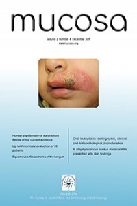
Results Of the 79 patients, 40 were male (50.6%) and 39 (49.4%) were female. Patients were aged between 18-91, with a mean overall age of 58.73 ± 17.95 years. Evaluation of the risk factors revealed that 45 patients (56.9%) were smokers, and alcohol was regularly consumed by 22 patients (27.8%). Most of the lesions were located in the buccal mucosa (34.4%). Homogenous OL was seen in 39 patients (49.4%) and nonhomogeneous OL in 40 patients (50.6%). The most reported histologic diagnosis was 37 (46.8%) cases of squamous hyperplasia (SH) and hyperkeratosis.
Conclusion To minimize the risk of malignant transformation in leukoplakia lesions, the elimination of risk factors and early biopsy is essential.
References
- 1. Brouns ER, Baart JA, Bloemena E, Karagozoglu H, van der Waal I. The relevance of uniform reporting in oral leukoplakia: definition, certainty factor and staging based on experience with 275 patients. Med Oral Patol Oral Cir Bucal 2013;18:19-26.
- 2. Petti S. Pooled estimate of world leukoplakia prevalence: a systematic review. Oral Oncology 2003;39:770-80.
- 3. Parlatescu I, Gheorghe C, Coculescu E, Tovaru S. Oral leukoplakia - an update. Maedica (Buchar) 2014;9:88-93.
- 4. Einhorn J, Wersall J. Incidence of oral carcinoma in patients with leukoplakia of the oral mucosa. Cancer 1967;20:2189-93.
- 5. Riedler L. ‘Bowenoid’ leukoplaki in the anal region. Klin Wochenschr 1988;66:271-3.
- 6. Morton TH, Cabay RJ, Epstein JB. Proliferative verrucous leukoplakia and its progression to oral carcinoma: report of three cases. J Oral Pathol Med 2007;36:315-8.
- 7. van der Waal I. Potentially malignant disorders of the oral and oropharyngeal mucosa; terminology, classification and present concepts of management. Oral Oncol 2009;45:317-23.
- 8. Özbayrak S. Ağız Hastalıkları Atlası. İstanbul: Quintessence Yayıncılık Ltd. Şti, 2003.
- 9. Neville B.W.,Damn D.D., Allen C.M., Bouquot J.E.: Oral & Maxillofacial Pathology. Saunders, Philadelphia 2002.
- 10. Axéll T,Pinborg J J, Smith C J, van der Waal I. Oral white lesions with special reference to precancerous and tobacco- related lesions: conclusions of an international symposium held in Uppsala, Sweden, May 18-21 1994. International Collaborative Group on Oral White Lesions. Oral Pathol Med 1994;25:49-54.
- 11. Holmstrup P, Vedtofte P, Reibel J, Stoltze K. Longterm treatment outcome of oral premalignant lesions. Oral Oncology 2006;42:461-74.
- 12. Bánóczy J, Squier C A, Kremer M, et al. The permeability of oral leukoplakia. Eur J Oral Sci, 2003;111:312-5.
- 13. Starzynska A, Pawlowska A, Renkielska D, Michajlowski I, Sobjanek M, Blazewicz I. Oral premalignant lesions: epidemiological and clinical analysis in the northern Polish population. Postepy Dermatol Alergol 2014;31:341-50.
- 14. Liu W, Wang YF, Zhou HW, Shi P, Zhou ZT, Tang GY. Malignant transformation of oral leukoplakia: a retrospective cohort study of 218 Chinese patients. BMC Cancer 2010;10:685.
- 15. Bisht RS, Singh AK, Sikarwar V, Darbari A. Study over the clinical picture and histopathology of leukoplakia and to establish the correlation between causative factors in the patients of Garhwal hill region. Natl J Maxillofac Surg 2013;4:177-80.
- 16. Lee JJ, Hung HC, Cheng SJ, et al. Carcinoma and dysplasia in oral leukoplakias in Taiwan: prevalence and risk factors. Oral Surg Oral Med Oral Pathol Oral Radiol Endod 2006;101:472-80.
- 17. Bouquot J E,Whitaker S B. Oral Leukoplakia -Rationale for diagnos is and prognosis of its clinical subtypes or ‘phases’. Quintessence Int 1994;25:133-40.
- 18. Sciubba JJ: Oral leukoplakia. Crit Rev Oral Biol Med 1995;6:147-60.
- 19. Dilhari A, Weerasekera MM, Siriwardhana A, et al. Candida infection in oral leukoplakia: an unperceived public health problem. Acta Odontol Scand 2016;74:565-9.
- 20. Sarkar R, Rathod GP. Clinicopathologic assessment of Candida colonization of oral leukoplakia. Indian J Dermatol Venereol Leprol 2014; 80:413-8.
- 21. Chandran R, Meer S, Feller L. Oral leukoplakia in a South African sample: a clinicopathological study. Oral Dis 2013;19:592-7.
Abstract
Amaç Oral lökoplaki (OL), oral mukozanın en yaygın prekanseröz lezyonudur. Etiyolojide en sık sorumlu tutulan faktörler, tütün ve alkol kullanımıdır. En yaygın yerleşim bölgesi bukkal mukoza olmakla birlikte, oral mukozanın diğer bölgelerini de tutabilir. Histopatolojik olarak lezyon spektrumu, displazinin eşlik etmediği skuamöz hiperplaziden; hafif, orta ve şiddetli displaziye kadar değişkenlik gösterebilir. Malign transformasyon riskinin en önemli belirleyicisi ise; displazi varlığı ile displazi derecesidir.
Yöntem Çalışmamızda, 2007-2015 yılları arasında Ege Üniversitesi Tıp Fakültesi yüz ve ağız lezyonları (EGEYA) konseyinde; klinik ve histopatolojik olarak oral lökoplaki tanısı alan hastalar, konsey bilgi formları ve fotoğraf arşivi üzerinden, hastaların demografik özellikleri de dikkate alınmak suretiyle, retrospektif olarak değerlendirildi.
Bulgular 79 hastanın 40’ı (%50.6) erkek ve 39’u (%9.4) kadındı. Hastalar 18 ile 91 yaşları arasında idi, ortalama yaş 58.73 ± 17.95 yıldı. Risk faktörleri açısından, 45 hasta (%56.9) sigara içiyor, 22 hasta (%27.8) düzenli alkol tüketiyordu. Lezyonların çoğu bukkal mukozadydı (%34.4). 39 hastada (%49.4) homojen, 40 hastada (%50.6) homojen olmayan OL saptandı. En fazla görülen histolojik tanı 37 hastada (%46.8) skuamöz hiperplazi (SH) ve hiperkeratozdu.
Sonuç Lökoplaki lezyonlarında malign transformasyon riskini en aza indirmek için risk faktörlerinin ortadan kaldırılması ve erken biyopsi alınması önemlidir.
References
- 1. Brouns ER, Baart JA, Bloemena E, Karagozoglu H, van der Waal I. The relevance of uniform reporting in oral leukoplakia: definition, certainty factor and staging based on experience with 275 patients. Med Oral Patol Oral Cir Bucal 2013;18:19-26.
- 2. Petti S. Pooled estimate of world leukoplakia prevalence: a systematic review. Oral Oncology 2003;39:770-80.
- 3. Parlatescu I, Gheorghe C, Coculescu E, Tovaru S. Oral leukoplakia - an update. Maedica (Buchar) 2014;9:88-93.
- 4. Einhorn J, Wersall J. Incidence of oral carcinoma in patients with leukoplakia of the oral mucosa. Cancer 1967;20:2189-93.
- 5. Riedler L. ‘Bowenoid’ leukoplaki in the anal region. Klin Wochenschr 1988;66:271-3.
- 6. Morton TH, Cabay RJ, Epstein JB. Proliferative verrucous leukoplakia and its progression to oral carcinoma: report of three cases. J Oral Pathol Med 2007;36:315-8.
- 7. van der Waal I. Potentially malignant disorders of the oral and oropharyngeal mucosa; terminology, classification and present concepts of management. Oral Oncol 2009;45:317-23.
- 8. Özbayrak S. Ağız Hastalıkları Atlası. İstanbul: Quintessence Yayıncılık Ltd. Şti, 2003.
- 9. Neville B.W.,Damn D.D., Allen C.M., Bouquot J.E.: Oral & Maxillofacial Pathology. Saunders, Philadelphia 2002.
- 10. Axéll T,Pinborg J J, Smith C J, van der Waal I. Oral white lesions with special reference to precancerous and tobacco- related lesions: conclusions of an international symposium held in Uppsala, Sweden, May 18-21 1994. International Collaborative Group on Oral White Lesions. Oral Pathol Med 1994;25:49-54.
- 11. Holmstrup P, Vedtofte P, Reibel J, Stoltze K. Longterm treatment outcome of oral premalignant lesions. Oral Oncology 2006;42:461-74.
- 12. Bánóczy J, Squier C A, Kremer M, et al. The permeability of oral leukoplakia. Eur J Oral Sci, 2003;111:312-5.
- 13. Starzynska A, Pawlowska A, Renkielska D, Michajlowski I, Sobjanek M, Blazewicz I. Oral premalignant lesions: epidemiological and clinical analysis in the northern Polish population. Postepy Dermatol Alergol 2014;31:341-50.
- 14. Liu W, Wang YF, Zhou HW, Shi P, Zhou ZT, Tang GY. Malignant transformation of oral leukoplakia: a retrospective cohort study of 218 Chinese patients. BMC Cancer 2010;10:685.
- 15. Bisht RS, Singh AK, Sikarwar V, Darbari A. Study over the clinical picture and histopathology of leukoplakia and to establish the correlation between causative factors in the patients of Garhwal hill region. Natl J Maxillofac Surg 2013;4:177-80.
- 16. Lee JJ, Hung HC, Cheng SJ, et al. Carcinoma and dysplasia in oral leukoplakias in Taiwan: prevalence and risk factors. Oral Surg Oral Med Oral Pathol Oral Radiol Endod 2006;101:472-80.
- 17. Bouquot J E,Whitaker S B. Oral Leukoplakia -Rationale for diagnos is and prognosis of its clinical subtypes or ‘phases’. Quintessence Int 1994;25:133-40.
- 18. Sciubba JJ: Oral leukoplakia. Crit Rev Oral Biol Med 1995;6:147-60.
- 19. Dilhari A, Weerasekera MM, Siriwardhana A, et al. Candida infection in oral leukoplakia: an unperceived public health problem. Acta Odontol Scand 2016;74:565-9.
- 20. Sarkar R, Rathod GP. Clinicopathologic assessment of Candida colonization of oral leukoplakia. Indian J Dermatol Venereol Leprol 2014; 80:413-8.
- 21. Chandran R, Meer S, Feller L. Oral leukoplakia in a South African sample: a clinicopathological study. Oral Dis 2013;19:592-7.
Details
| Primary Language | English |
|---|---|
| Subjects | Clinical Sciences |
| Journal Section | Original Articles |
| Authors | |
| Publication Date | December 29, 2019 |
| Published in Issue | Year 2019 Volume: 2 Issue: 4 |