Öz
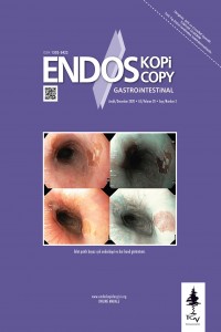
Giriş ve Amaç: İnlet patch, üst özofagus sfinkterinde veya hemen distalinde yer alan heterotopik gastrik mukoza adasıdır. Bu çalışmada amaç kliniğimizde inlet patch tanısı konulan vakaların sıklığı, demografik, klinik ve endoskopik özelliklerini değerlendirmektir. Gereç ve Yöntem: Bu çalışma Ocak 2015- Mart 2020 tarihleri arasında Gastroenteroloji Bilim Dalında herhangi bir nedenle özofagogastroduodenoskopi yapılıp, inlet patch tanısı konulan 245 hastanın retrospektif değerlendirilmesini içermektedir. Çalışmaya alınan hastaların; yaş, cinsiyet, endoskopi yapılma nedeni, inlet patch lezyonunun boyutu ve sayısı, Barrett özofagus, özofajit ve hiatus hernisi varlığı ve var olan patoloji sonuçları değerlendirilmiştir. Bulgular: İki yüz kırk beş hastada inlet patch bulunmuştur. İki yüz kırk beş hastanın 124’ü (%50.6) kadın, yaş ortalaması 48.64±14.54 yıldır. İnlet patch boyutunun ortalaması 13.32±8.85 (3-40) mm’dir. En sık endoskopi yapılma nedeni 91 (%37.1) hastada dispepsi olarak saptanmıştır. İnlet patch saptanan hastaların endoskopi sırasında 39’unda (%15.9) özofajit, 20’sinde (%8.2) hiatus hernisi ve 6’sında (%2.4) Barrett özofagus görülmüştür. Hastaların 125’inden (%51) biyopsi alınmış olup, 98 (%78.4) hastada patoloji ile uyumlu sonuçlanmıştır. Hastaların endoskopi yapılma nedenleri, Barrett özofagus ve hiatus hernisi varlığı ile inlet patch boyutu arasındaki istatistiksel olarak anlamlı farklılık olduğu saptanmıştır (sırasıyla; p=0.03, p=0.004, p=0016). Sonuç: Herhangi bir nedenle yapılan endoskopilerin %1.24’ünde inlet patch saptanmıştır. Merkezimiz üçüncü basamak bir sağlık kuruluşu olduğundan bu sonucun, Ege Bölgesi’nin inlet patch prevalansını yansıttığını düşünmekteyiz. Fonksiyonel dispepsi, disfaji, nedeni bilinmeyen kronik öksürüğü ve globusu olan hastalarda, servikal özofagus inlet patch açısından dikkatli bir şekilde incelenmelidir.
Anahtar Kelimeler
Kaynakça
- 1. Schumidt FFA. De mammlium oesophage atque ventriculo, Inaugural dissertation. Bathenea: Halle, 1805.
- 2. Maconi G, Pace F, Vago L, et al. Prevelance and clinical features of heterotopic gastric mucosa in the upper oesophagus (inlet patch). Eur J Gastroenterol Hepatol. 2000;12:745-9.
- 3. Peitz U, Vieth M, Evert M, et al. The prevalence of gastric heterotopia of the proximal esophagus is underestimated, but preneoplasia is rare—correlation with Barrett’s esophagus. BMC Gastroenterol 2017;17:87.
- 4. Jacobs E, Dehou MF. Heterotopic gastric mucosa in the upper esophagus: a prospective study of 33 cases and review of literature. Endoscopy 1997;29:710-5.
- 5. Lujan G, Genta R. The inlet patch revisited: A clinicopathologic study of 569 patients with heterotopic gastric mucosa in the proximal esophagus. Am J Gastroenterol. 2010;105:S4.
- 6. Weickert U, Wolf A, Schröder C, et al. Frequency, histopathological findings, and clinical significance of cervical heterotopic gastric mucosa (gastric inlet patch): a prospective study in 300 patients. Dis Esophagus 2011;24:63-8.
- 7. Rector LE, Connerley ML. Aberrant mucosa in esophagus in infants and in children. Arch Pathol 1941;31:285.
- 8. Jabbari M, Goresky CA, Lough J, et al. The inlet patch: heterotopic gastric mucosa in the upper esophagus. Gastroenterology 1985;89:352-6.
- 9. Tang P, McKinley MJ, Sporrer M, Kahn E. Inlet patch: prevalence, histologic type, and association with esophagitis, Barrett esophagus, and antritis. Arch Pathol Lab Med 2004;128:444-7.
- 10. Shah KK, DeRidder PH, Shah KK. Ectopic gastric mucosa in proximal esophagus. Its clinical significance and hormonal profile. J Clin Gastroenterol 1986;8:509-13.
- 11. Rusu R, Ishaq S, Wong T, Dunn JM. Cervical inlet patch: new insights into diagnosis and endoscopic therapy. Frontline Gastroenterol 2018;9:214-20.
- 12. American Gastroenterological Association, Spechler SJ, Sharma P, Souza RF, Inadomi JM, Shaheen NJ. American Gastroenterological Association medical position statement on the management of Barrett's esophagus. Gastroenterology 2011;140:1084-91.
- 13. Lundell LR, Dent J, Bennett JR, et al. Endoscopic assessment of oesophagitis: clinical and functional correlates and further validation of the Los Angeles classification. Gut 1999;45:172-80.
- 14. Al-Mammari S, Selvarajah U, East JE, Bailey AA, Braden B. Narrow band imaging facilitates detection of inlet patches in the cervical oesophagus. Dig Liver Dis 2014;46:716-9.
- 15. Kekilli M, Sayılır M, Yeşil Y, et al. Servikal özofagustaki HGM’nın endoskopik sıklığı; bir referans merkez çalışması. Akademik Gastroenteroloji Dergisi 2009;8:119-22.
- 16. Akbayır N, Alkim C, Erdem L, et al. Heterotopic gastric mucosa in the servical esophagus (inlet patch): endoscopic prevalence, histological and clinical characteristics. J Gastroenterol Hepatol 2004;19:891-6.
- 17. Yüksel I, Üsküdar O, Köklü S, et al. Inlet patch: Association with endoscopic findings in the upper gastrointestinal system. Scand J Gastroenterol 2008;43:910-4.
- 18. Savaş N, Akbaş E. Heterotopik gastrik mukozanın sıklığı, klinik önemi ve eşlik eden diğer klinik bulgular. Endoskopi Gastrointestinal 2014;22:60-3.
- 19. Poyrazoğlu OK, Bahçecioğlu IH, Dağlı AF, et al. Heterotopic gastric mucosa (inlet patch): endoscopic prevalence, histopathological, demographical and clinical characteristics. Int J Clin Pract 2009;63:287-91.
- 20. Takeji H, Ueno J, Nishitani H. Ectopic gastric mucosa in the upper esophagus: prevalence and radiologic findings. AJR Am J Roentgenol 1995;164:901-4.
- 21. Alagozlu H, Simsek Z, Unal S, et al. Is there an association between Helicobacter pylori in the inlet patch and globus sensation? World J Gastroenterol 2010;16:42-7.
- 22. Ciocalteu A, Popa P, Ionescu M, Gheonea DI. Issues and cont.roversies in esophageal inlet patch. World J Gastroenterol 2019;25:4061-73.
- 23. Neumann WL, Luján GM, Genta RM. Gastric heterotopia in the proximal oesophagus ("inlet patch"): Association with adenocarcinomas arising in Barrett mucosa. Dig Liver Dis 2012;44:292-6.
- 24. von Rahden BH, Stein HJ, Becker K, Liebermann-Meffert D, Siewert JR. Heterotopic gastric mucosa of the esophagus: literature-review and proposal of a clinicopathologic classification. Am J Gastroenterol 2004;99:543-51.
- 25. Chong VH, Jalihal A. Heterotopic gastric mucosal patch of the esophagus is associated with higher prevalence of laryngopharyngeal reflux symptoms. Eur Arch Otorhinolaryngol 2010;267:1793-9.
- 26. Bajbouj M, Becker V, Eckel F, et al. Argon plasma coagulation of cervical heterotopic gastric mucosa as an alternative treatment for globus sensations. Gastroenterology 2009;137:440-4.
- 27. López-Colombo A, Jiménez-Toxqui M, Gogeascoechea-Guillén PD, et al. Prevalence of esophageal inlet patch and clinical characteristics of the patients. Rev Gastroenterol Mex 2019;84:442-8.
- 28. Lauwers GY, Mino M, Ban S, et al. Cytokeratins 7 and 20 and mucin core protein expression in esophageal cervical inlet patch. Am J Surg Pathol 2005;29:437-42.
- 29. Avidan B, Sonnenberg A, Chejfec G, Schnell TG, Sontag SJ. Is there a link between cervical inlet patch and Barrett's esophagus? Gastrointest Endosc 2001;53:717-21.
- 30. Borhan-Manesh F, Farnum JB. Incidence of heterotopic gastric mucosa in the upper oesophagus. Gut 1991;32:968-72.
- 31. Behrens C, Yen PP. Esophageal inlet patch. Radiol Res Pract 2011;2011:460890.
- 32. Shimamura Y, Winer S, Marcon N. A giant circumferential inlet patch with acid secretion causing stricture. Clin Gastroenterol Hepatol 2017;15:A22-A23.
- 33. Hori K, Kim Y, Sakurai J, et al. Non-erosive reflux disease rather than cervical inlet patch involves globus. J Gastroenterol 2010;45:1138-45.
Öz
Background and Aims: An inlet patch is an island of heterotopic gastric mucosa located in the upper or immediate distal part of the esophageal sphincter. The aim of this study was to evaluate the frequency, demographics, and clinical and endoscopic features of cases diagnosed with inlet patch in our clinic. Material and Methods: This retrospective study included 245 patients who underwent esophagogastroduodenoscopy for any reason in the department of gastroenterology between January 2015 and March 2020. The patients were evaluated on the basis of age, gender, reason for endoscopy, size and number of inlet patch lesions, presence of Barrett’s esophagus, esophagitis and hiatus hernia, and pathology results, if available. Results: İnlet patch was found in 245 of the endoscopies performed for any reason. Of the 245 patients, 124 (50.6%) were women, and the mean age was 48.64±14.54 (19-81) years. The mean size of inlet patch was 13.32±8.85 (3-40) mm. Dyspepsia was found to be the most common reason for endoscopy in 91 (37.1%) patients. Endoscopy revealed esophagitis in 39 (15.9%), hiatus hernias in 20 (8.2%), and Barrett’s esophagus in 6 (2.4%) patients among those detected to have inlet patch. A biopsy was taken from 125 (51%) patients, and the result was consistent with the reported pathology in 98 (78.4%) patients. A statistically significant difference was found between the inlet patch size and the reason for endoscopy, presence of Barrett’s esophagus, and presence of hiatus hernia (p=0.03, 0.004, and 0.016, respectively). Conclusion: İnlet patch was detected in 1.24% of endoscopies performed. Since ours is tertiary healthcare providing center, we consider that this result reflects the inlet patch prevalence of the Ege region in Turkey. The cervical esophagus should be carefully examined for the possibility of inlet patch in patients with functional dyspepsia, dysphagia, chronic cough with an unknown cause, and globus. Endoscopy should be repeated using narrow-band imaging technique after fully sedating the patients even if endoscopy has been performed before.
Anahtar Kelimeler
Kaynakça
- 1. Schumidt FFA. De mammlium oesophage atque ventriculo, Inaugural dissertation. Bathenea: Halle, 1805.
- 2. Maconi G, Pace F, Vago L, et al. Prevelance and clinical features of heterotopic gastric mucosa in the upper oesophagus (inlet patch). Eur J Gastroenterol Hepatol. 2000;12:745-9.
- 3. Peitz U, Vieth M, Evert M, et al. The prevalence of gastric heterotopia of the proximal esophagus is underestimated, but preneoplasia is rare—correlation with Barrett’s esophagus. BMC Gastroenterol 2017;17:87.
- 4. Jacobs E, Dehou MF. Heterotopic gastric mucosa in the upper esophagus: a prospective study of 33 cases and review of literature. Endoscopy 1997;29:710-5.
- 5. Lujan G, Genta R. The inlet patch revisited: A clinicopathologic study of 569 patients with heterotopic gastric mucosa in the proximal esophagus. Am J Gastroenterol. 2010;105:S4.
- 6. Weickert U, Wolf A, Schröder C, et al. Frequency, histopathological findings, and clinical significance of cervical heterotopic gastric mucosa (gastric inlet patch): a prospective study in 300 patients. Dis Esophagus 2011;24:63-8.
- 7. Rector LE, Connerley ML. Aberrant mucosa in esophagus in infants and in children. Arch Pathol 1941;31:285.
- 8. Jabbari M, Goresky CA, Lough J, et al. The inlet patch: heterotopic gastric mucosa in the upper esophagus. Gastroenterology 1985;89:352-6.
- 9. Tang P, McKinley MJ, Sporrer M, Kahn E. Inlet patch: prevalence, histologic type, and association with esophagitis, Barrett esophagus, and antritis. Arch Pathol Lab Med 2004;128:444-7.
- 10. Shah KK, DeRidder PH, Shah KK. Ectopic gastric mucosa in proximal esophagus. Its clinical significance and hormonal profile. J Clin Gastroenterol 1986;8:509-13.
- 11. Rusu R, Ishaq S, Wong T, Dunn JM. Cervical inlet patch: new insights into diagnosis and endoscopic therapy. Frontline Gastroenterol 2018;9:214-20.
- 12. American Gastroenterological Association, Spechler SJ, Sharma P, Souza RF, Inadomi JM, Shaheen NJ. American Gastroenterological Association medical position statement on the management of Barrett's esophagus. Gastroenterology 2011;140:1084-91.
- 13. Lundell LR, Dent J, Bennett JR, et al. Endoscopic assessment of oesophagitis: clinical and functional correlates and further validation of the Los Angeles classification. Gut 1999;45:172-80.
- 14. Al-Mammari S, Selvarajah U, East JE, Bailey AA, Braden B. Narrow band imaging facilitates detection of inlet patches in the cervical oesophagus. Dig Liver Dis 2014;46:716-9.
- 15. Kekilli M, Sayılır M, Yeşil Y, et al. Servikal özofagustaki HGM’nın endoskopik sıklığı; bir referans merkez çalışması. Akademik Gastroenteroloji Dergisi 2009;8:119-22.
- 16. Akbayır N, Alkim C, Erdem L, et al. Heterotopic gastric mucosa in the servical esophagus (inlet patch): endoscopic prevalence, histological and clinical characteristics. J Gastroenterol Hepatol 2004;19:891-6.
- 17. Yüksel I, Üsküdar O, Köklü S, et al. Inlet patch: Association with endoscopic findings in the upper gastrointestinal system. Scand J Gastroenterol 2008;43:910-4.
- 18. Savaş N, Akbaş E. Heterotopik gastrik mukozanın sıklığı, klinik önemi ve eşlik eden diğer klinik bulgular. Endoskopi Gastrointestinal 2014;22:60-3.
- 19. Poyrazoğlu OK, Bahçecioğlu IH, Dağlı AF, et al. Heterotopic gastric mucosa (inlet patch): endoscopic prevalence, histopathological, demographical and clinical characteristics. Int J Clin Pract 2009;63:287-91.
- 20. Takeji H, Ueno J, Nishitani H. Ectopic gastric mucosa in the upper esophagus: prevalence and radiologic findings. AJR Am J Roentgenol 1995;164:901-4.
- 21. Alagozlu H, Simsek Z, Unal S, et al. Is there an association between Helicobacter pylori in the inlet patch and globus sensation? World J Gastroenterol 2010;16:42-7.
- 22. Ciocalteu A, Popa P, Ionescu M, Gheonea DI. Issues and cont.roversies in esophageal inlet patch. World J Gastroenterol 2019;25:4061-73.
- 23. Neumann WL, Luján GM, Genta RM. Gastric heterotopia in the proximal oesophagus ("inlet patch"): Association with adenocarcinomas arising in Barrett mucosa. Dig Liver Dis 2012;44:292-6.
- 24. von Rahden BH, Stein HJ, Becker K, Liebermann-Meffert D, Siewert JR. Heterotopic gastric mucosa of the esophagus: literature-review and proposal of a clinicopathologic classification. Am J Gastroenterol 2004;99:543-51.
- 25. Chong VH, Jalihal A. Heterotopic gastric mucosal patch of the esophagus is associated with higher prevalence of laryngopharyngeal reflux symptoms. Eur Arch Otorhinolaryngol 2010;267:1793-9.
- 26. Bajbouj M, Becker V, Eckel F, et al. Argon plasma coagulation of cervical heterotopic gastric mucosa as an alternative treatment for globus sensations. Gastroenterology 2009;137:440-4.
- 27. López-Colombo A, Jiménez-Toxqui M, Gogeascoechea-Guillén PD, et al. Prevalence of esophageal inlet patch and clinical characteristics of the patients. Rev Gastroenterol Mex 2019;84:442-8.
- 28. Lauwers GY, Mino M, Ban S, et al. Cytokeratins 7 and 20 and mucin core protein expression in esophageal cervical inlet patch. Am J Surg Pathol 2005;29:437-42.
- 29. Avidan B, Sonnenberg A, Chejfec G, Schnell TG, Sontag SJ. Is there a link between cervical inlet patch and Barrett's esophagus? Gastrointest Endosc 2001;53:717-21.
- 30. Borhan-Manesh F, Farnum JB. Incidence of heterotopic gastric mucosa in the upper oesophagus. Gut 1991;32:968-72.
- 31. Behrens C, Yen PP. Esophageal inlet patch. Radiol Res Pract 2011;2011:460890.
- 32. Shimamura Y, Winer S, Marcon N. A giant circumferential inlet patch with acid secretion causing stricture. Clin Gastroenterol Hepatol 2017;15:A22-A23.
- 33. Hori K, Kim Y, Sakurai J, et al. Non-erosive reflux disease rather than cervical inlet patch involves globus. J Gastroenterol 2010;45:1138-45.
Ayrıntılar
| Birincil Dil | Türkçe |
|---|---|
| Konular | Sağlık Kurumları Yönetimi |
| Bölüm | Makaleler |
| Yazarlar | |
| Yayımlanma Tarihi | 30 Aralık 2020 |
| Yayımlandığı Sayı | Yıl 2020 Cilt: 28 Sayı: Sayı: 3 |


