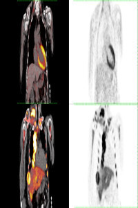
Hypermetabolic Gall Bladder Wall Thickening Mimicking Benign Process: Adenomyomatosis
Abstract
Keywords
References
- Oe, A., Kawabe, J., Torii, K., Kawamura, E., Higashiyama, S., Kotani, J., Hayashi, T., Kurooka, H., Tsumoto, C., Kubo, S., & Shiomi, S. (2006). Distinguishing benign from malignant gallbladder wall thickening using FDG-PET. Annals of nuclear medicine, 20(10), 699–703. https://doi.org/10.1007/BF02984683
- Gupta, V., Vishnu, K. S., Yadav, T. D., Sakaray, Y. R., Irrinki, S., Mittal, B. R., Kalra, N., & Vaiphei, K. (2019). Radio-pathological Correlation of 18F-FDG PET in Characterizing Gallbladder Wall Thickening. Journal of gastrointestinal cancer, 50(4), 901–906. https://doi.org/10.1007/s12029-018-0176-2
- Koh, T., Taniguchi, H., Kunishima, S., & Yamagishi, H. (2000). Possibility of Differential Diagnosis of Small Polypoid Lesions in the Gallbladder Using FDG-PET. Clinical positron imaging : official journal of the Institute for Clinical P.E.T, 3(5), 213–218. https://doi.org/10.1016/s1095-0397(00)00100-x
- Maldjian, P. D., Ghesani, N., Ahmed, S., & Liu, Y. (2007). Adenomyomatosis of the gallbladder: another cause for a "hot" gallbladder on 18F-FDG PET. AJR. American journal of roentgenology, 189(1), W36–W38. https://doi.org/10.2214/AJR.05.1284
- Yoshimitsu, K., Honda, H., Aibe, H., Shinozaki, K., Kuroiwa, T., Irie, H., Asayama, Y., & Masuda, K. (2001). Radiologic diagnosis of adenomyomatosis of the gallbladder: comparative study among MRI, helical CT, and transabdominal US. Journal of computer assisted tomography, 25(6), 843–850. https://doi.org/10.1097/00004728-200111000-00003
- Tomizawa, M., Shinozaki, F., Uchida, Y., Uchiyama, K., Tanaka, S., Sunaoshi, T., Kano, D., Sugiyama, E., Shite, M., Haga, R., Fukamizu, Y., Fujita, T., Kagayama, S., Hasegawa, R., Shirai, Y., Motoyoshi, Y., Sugiyama, T., Yamamoto, S., & Ishige, N. (2017). Comparison of DWIBS/T2 image fusion and PET/CT for the diagnosis of cancer in the abdominal cavity. Experimental and therapeutic medicine, 14(4), 3754–3760. https://doi.org/10.3892/etm.2017.4987
- Joo, I., Lee, J. Y., Kim, J. H., Kim, S. J., Kim, M. A., Han, J. K., & Choi, B. I. (2013). Differentiation of adenomyomatosis of the gallbladder from early-stage, wall-thickening-type gallbladder cancer using high-resolution ultrasound. European radiology, 23(3), 730–738. https://doi.org/10.1007/s00330-012-2641-9
- Suzuki, K., Watada, S., Yoko, M., Nakahara, T., & Kumamoto, Y. (2011). Successful diagnosis of gallbladder carcinoma coexisting with adenomyomatosis by 18F-FDG-PET--report of a case. Journal of gastrointestinal cancer, 42(4), 252–256. https://doi.org/10.1007/s12029-010-9221-5
Details
Primary Language
English
Subjects
Gastroenterology and Hepatology, Nuclear Medicine, Pathology, Radiology and Organ Imaging
Journal Section
Case Report
Authors
Zehra Pınar Koç
*
0000-0002-3274-5790
Türkiye
Pınar Pelin Özcan
0000-0003-0147-2678
Türkiye
F. Demir Apaydın
0000-0001-7023-4521
Türkiye
Esra Zeynep Coşkunoğlu
This is me
0000-0002-6176-7016
Türkiye
Publication Date
May 14, 2024
Submission Date
April 4, 2024
Acceptance Date
May 14, 2024
Published in Issue
Year 2024 Volume: 4 Number: 1