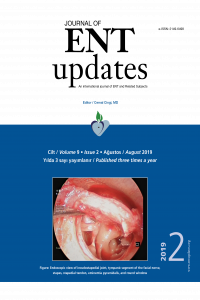
The relationship between superior attachment of the uncinate process of the ethmoid and varying paranasal sinus anatomy: an analysis using computerised tomography
Öz
Anahtar Kelimeler
Paranasal sinuses uncinate process turbinates skull base multidetector computed tomography
Kaynakça
- 1. Başak S, Akdilli A, Karaman CZ, Kunt T. Assessment of some important anatomical variations and dangerous areas of the paranasal sinuses by computed tomography in children. Int J Pediatr Otorhinolaryngol 2000;55:81-9.
- 2. Wormald PJ. The agger nasi cell: the key to understanding the anatomy of the frontal recess. Otolaryngol Head Neck Surg 2003;129:497-507.
- 3. Netto B, Piltcher OB, Meotti CD, Lemieszek J, Isolan GR. Computed tomography imaging study of the superior attachment of the uncinate process. Rhinology 2015;53:187-91.
- 4. Tuli IP, Sengupta S, Munjal S, Kesari SP, Chakraborty S. Anatomical variations of uncinate process observed in chronic sinusitis. Indian J Otolaryngol Head Neck Surg 2013;65:157-61.
- 5. Mahmutoğlu AS, Çelebi I, Akdana B, et al. Computed tomographic analysis of frontal sinus drainage pathway variations and frontal rhinosinusitis. J Craniofac Surg 2015;26:87-90.
- 6. Landsberg R, Friedman M. A computer assisted anatomical study of the nasofrontal region. Laryngoscope 2001;111:2125-30.
- 7. Srivastava M, Sushant T. Role of anatomic variations of uncinate process in frontal sinusitis. Indian J Otolaryngol Head Neck Surg 2016;68:441-4.
- 8. Stammberger H, Hawke M. Functional endoscopic sinus surgery: the Messerklinger technique. Decker, Philadelphia, 1999 pp. 61-90.
- 9. Badia L, Lund VJ, Wei W, Ho WK. Ethnic variation in sinonasal anatomy on CT-scanning. Rhinology 2005;43:210-4.
- 10. Güldner C, Zimmermann AP, Diogo I, Werner JA, Teymoortash A. Age dependent difference of the anterior skull base Int J Pediatr Otorhinolaryngol 2012;76:822-8.
- 11. Keros P. Uber die praktische bedeutung der niveauunterschiede der lamina cribrosa des etmoids. [Article in Germany] Z Lar Rhin Otol 1965;41:808-10.
- 12. Bingham B, Wang RG, Hawke M, Kwok P. The embryonic development of the lateral nasal wall from 8 to 24 weeks. Laryngoscope 1991;101:992-7.
- 13. Nayak DR, Balakrishnan R, Murty KD. Functional anatomy of the uncinate process and its role in endoscopic sinus surgery. Indian J Otolaryngol Head Neck Surg 2001;53:27-31.
- 14. Lund VJ, Stammberger H, Fokkens WJ, et al. European position paper on the anatomical terminology of the internal nose and paranasal sinuses. Rhinol Suppl 2014;24:1-34.
- 15. Wake M, Takeno S, Hawke M. The uncinate process: a histological and morphological study. Laryngoscope 1994;104:364-9.
- 16. Cheng SY, Yang CJ, Lee CH, et al. The association of superior attachment of uncinate process with pneumatization of middle turbinate: a computed tomographic analysis. Eur Arch Otorhinolaryngol 2017; 274: 1905-10.
- 17. Liu SC, Wang CH, Wang HW. Prevalence of the uncinate process, agger nasi cell and relationship in a Taiwanese population. Rhinology 2010;48:239-44.
- 18. Ercan I, Cakır BO, Sayın I, Başak M, Turgut S. Relationship between the superior attachments type of uncinate process and presence of agger nasi cell: a computer-assisted anatomic study. Otolaryngol Head Neck Surg 2006;134:1010-4.
- 19. Messerklinger W. On the drainage of the normal frontal sinus of man. Acta Otolaryngol (Stockh) 1967; 63:176-81.
- 20. Kantarci M, Karasen RM, Alper F, Onbas O, Okur A, Karaman A. Remarkable anatomic variations in paranasal sinus region and their clinical importance. Eur J Radiol 2004;50:296-302.
- 21. Fadda GL, Rosso S, Aversa S, Petrelli A, Ondolo C, Succo G. Multiparametric statistical correlations between paranasal sinus anatomic variations and chronic rhinosinusitis. Acta Otorhinolaryngol Ital 2012;32:244-51.
- 22. Krzeski A, Tomaszewska E, Jakubczyk I, Galewicz-Zielinska A. Anatomic variations of the lateral nasal wall in the computed tomography scans of patients with chronic rhinosinusitis. Am J Rhinol 2001;15:371-5.
- 23. Bolger WE, Butzin CA, Parsons DS. Paranasal sinus bony anatomic variation and mucosal abnormalities: CT analysis for endoscopic sinus surgery. Laryngoscope 1991;101:56-64.
- 24. Zhang L, Han D, Ge W, et al. Anatomical and computed tomographic analysis of the interaction between the uncinate process and the agger nasi cell. Acta Otolaryngol 2006;126:845-52.
- 25. Ozcan KM, Selcuk A, Ozcan I, Akdogan O, Dere H. Anatomical variations of nasal turbinates. J Craniofac Surg 2008;19:1678-82.
- 26. Gun R, Yorgancilar E, Bakir S, et al. The relationship between pneumatized middle turbinate and the anterior ethmoid roof dimensions: a radiologic study. Eur Arch Otorhinolaryngol 2013;270:1365-71.
- 27. Şahin C, Yılmaz YF, Titiz A, Ozcan M, Ozlügedik S, Unal A. Analysis of Ethmoid Roof and Cranial Base in Turkish Population. [Article in Turkish] KBBve BBC Derg 2007;15:1-6.
- 28. Gauba V, Saleh GM, Dua G, Agarwal S, Ell S, Vize C. Radiological classification of anterior skull base anatomy prior to performing medial orbital wall decompression. Orbit 2006;25:93-6.
- 29. Adeel M, Ikram M, Rajput MS, Arain A, Khattak YJ. Asymmetry of lateral lamella of the cribriform plate: a software-based analysis of coronal computed tomography and its clinical relevance in endoscopic sinus surgery. Surg Radiol Anat 201335:843-7.
Öz
Kaynakça
- 1. Başak S, Akdilli A, Karaman CZ, Kunt T. Assessment of some important anatomical variations and dangerous areas of the paranasal sinuses by computed tomography in children. Int J Pediatr Otorhinolaryngol 2000;55:81-9.
- 2. Wormald PJ. The agger nasi cell: the key to understanding the anatomy of the frontal recess. Otolaryngol Head Neck Surg 2003;129:497-507.
- 3. Netto B, Piltcher OB, Meotti CD, Lemieszek J, Isolan GR. Computed tomography imaging study of the superior attachment of the uncinate process. Rhinology 2015;53:187-91.
- 4. Tuli IP, Sengupta S, Munjal S, Kesari SP, Chakraborty S. Anatomical variations of uncinate process observed in chronic sinusitis. Indian J Otolaryngol Head Neck Surg 2013;65:157-61.
- 5. Mahmutoğlu AS, Çelebi I, Akdana B, et al. Computed tomographic analysis of frontal sinus drainage pathway variations and frontal rhinosinusitis. J Craniofac Surg 2015;26:87-90.
- 6. Landsberg R, Friedman M. A computer assisted anatomical study of the nasofrontal region. Laryngoscope 2001;111:2125-30.
- 7. Srivastava M, Sushant T. Role of anatomic variations of uncinate process in frontal sinusitis. Indian J Otolaryngol Head Neck Surg 2016;68:441-4.
- 8. Stammberger H, Hawke M. Functional endoscopic sinus surgery: the Messerklinger technique. Decker, Philadelphia, 1999 pp. 61-90.
- 9. Badia L, Lund VJ, Wei W, Ho WK. Ethnic variation in sinonasal anatomy on CT-scanning. Rhinology 2005;43:210-4.
- 10. Güldner C, Zimmermann AP, Diogo I, Werner JA, Teymoortash A. Age dependent difference of the anterior skull base Int J Pediatr Otorhinolaryngol 2012;76:822-8.
- 11. Keros P. Uber die praktische bedeutung der niveauunterschiede der lamina cribrosa des etmoids. [Article in Germany] Z Lar Rhin Otol 1965;41:808-10.
- 12. Bingham B, Wang RG, Hawke M, Kwok P. The embryonic development of the lateral nasal wall from 8 to 24 weeks. Laryngoscope 1991;101:992-7.
- 13. Nayak DR, Balakrishnan R, Murty KD. Functional anatomy of the uncinate process and its role in endoscopic sinus surgery. Indian J Otolaryngol Head Neck Surg 2001;53:27-31.
- 14. Lund VJ, Stammberger H, Fokkens WJ, et al. European position paper on the anatomical terminology of the internal nose and paranasal sinuses. Rhinol Suppl 2014;24:1-34.
- 15. Wake M, Takeno S, Hawke M. The uncinate process: a histological and morphological study. Laryngoscope 1994;104:364-9.
- 16. Cheng SY, Yang CJ, Lee CH, et al. The association of superior attachment of uncinate process with pneumatization of middle turbinate: a computed tomographic analysis. Eur Arch Otorhinolaryngol 2017; 274: 1905-10.
- 17. Liu SC, Wang CH, Wang HW. Prevalence of the uncinate process, agger nasi cell and relationship in a Taiwanese population. Rhinology 2010;48:239-44.
- 18. Ercan I, Cakır BO, Sayın I, Başak M, Turgut S. Relationship between the superior attachments type of uncinate process and presence of agger nasi cell: a computer-assisted anatomic study. Otolaryngol Head Neck Surg 2006;134:1010-4.
- 19. Messerklinger W. On the drainage of the normal frontal sinus of man. Acta Otolaryngol (Stockh) 1967; 63:176-81.
- 20. Kantarci M, Karasen RM, Alper F, Onbas O, Okur A, Karaman A. Remarkable anatomic variations in paranasal sinus region and their clinical importance. Eur J Radiol 2004;50:296-302.
- 21. Fadda GL, Rosso S, Aversa S, Petrelli A, Ondolo C, Succo G. Multiparametric statistical correlations between paranasal sinus anatomic variations and chronic rhinosinusitis. Acta Otorhinolaryngol Ital 2012;32:244-51.
- 22. Krzeski A, Tomaszewska E, Jakubczyk I, Galewicz-Zielinska A. Anatomic variations of the lateral nasal wall in the computed tomography scans of patients with chronic rhinosinusitis. Am J Rhinol 2001;15:371-5.
- 23. Bolger WE, Butzin CA, Parsons DS. Paranasal sinus bony anatomic variation and mucosal abnormalities: CT analysis for endoscopic sinus surgery. Laryngoscope 1991;101:56-64.
- 24. Zhang L, Han D, Ge W, et al. Anatomical and computed tomographic analysis of the interaction between the uncinate process and the agger nasi cell. Acta Otolaryngol 2006;126:845-52.
- 25. Ozcan KM, Selcuk A, Ozcan I, Akdogan O, Dere H. Anatomical variations of nasal turbinates. J Craniofac Surg 2008;19:1678-82.
- 26. Gun R, Yorgancilar E, Bakir S, et al. The relationship between pneumatized middle turbinate and the anterior ethmoid roof dimensions: a radiologic study. Eur Arch Otorhinolaryngol 2013;270:1365-71.
- 27. Şahin C, Yılmaz YF, Titiz A, Ozcan M, Ozlügedik S, Unal A. Analysis of Ethmoid Roof and Cranial Base in Turkish Population. [Article in Turkish] KBBve BBC Derg 2007;15:1-6.
- 28. Gauba V, Saleh GM, Dua G, Agarwal S, Ell S, Vize C. Radiological classification of anterior skull base anatomy prior to performing medial orbital wall decompression. Orbit 2006;25:93-6.
- 29. Adeel M, Ikram M, Rajput MS, Arain A, Khattak YJ. Asymmetry of lateral lamella of the cribriform plate: a software-based analysis of coronal computed tomography and its clinical relevance in endoscopic sinus surgery. Surg Radiol Anat 201335:843-7.
Ayrıntılar
| Birincil Dil | İngilizce |
|---|---|
| Konular | Sağlık Kurumları Yönetimi |
| Bölüm | Makaleler |
| Yazarlar | |
| Yayımlanma Tarihi | 8 Ağustos 2019 |
| Gönderilme Tarihi | 7 Haziran 2019 |
| Kabul Tarihi | 3 Temmuz 2019 |
| Yayımlandığı Sayı | Yıl 2019 Cilt: 9 Sayı: 2 |

