Öz
The present study was carried out to determine the plexus lumbosacralis and its branches in pigeon (Columba livia). In this study, 15 doves that were collected from Erzurum and its vicinity were used. First, 5 mg/kg xylazine and then 30 mg/kg ketalar were injected into the muscle of all materials for anesthesia. Then, blood was drained by opening the body cavity and the animals were fixed with formaldehyde. The nerves of plexus lumbosacralis were dissected separately and photographed. Plexus lumbosacralis was formed by the union of the branches of the synsacral spinal nerves leaving from the ventrolaterale of os lumbosacrale. The plexus consisted of seven (2-‐8.) nerves in pigeon (Columba livia). The nerves originating from the plexus lumbalis from the cranial to caudal were n. ilioinguinalis, n. cutaneus femoris, n. coxalis cranialis, n. femoralis, n. saphenus and n. obturatorius. Nervus coxalis caudalis, the common root of n. peroneus and n. tibialis, n. cutaneus femoris caudalis and the common branches of rr. musculares originated from the plexus sacralis. It was determined that general macroanatomical shapes of plexus lumbosacralis and the distribution of nerves originating from this plexus were found to be similar with a large extent in dove, as one of the wild bird species. Key words: Columba livia, Pigeon, Plexus lumbosacralis.
Anahtar Kelimeler
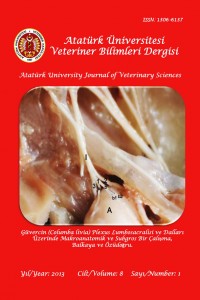
Güvercin (Columba livia) Plexus Lumbosacralisi ve Dalları Üzerinde Makroanatomik ve Subgros Bir Çalışma
Öz
Çalışma, güvercinin (Columba livia) plexus lumbosacralis’inin oluşumu ve plexus’tan ayrılan sinir dallarının belirlenmesi amacıyla yapıldı. Araştırmada materyal olarak Erzurum ve yöresinden toplanan 15 adet güvercin kullanıldı. Materyallere, anestezi için önce 5 mg/kg xylazine, sonra 30 mg/kg ketalar kas içine enjekte edildi. Anesteziyi takiben hayvanların vücut boşluğu açılarak kanı boşaltıldı ve formaldehit ile tespit edildi. Plexus lumbosacralis’i oluşturan ve plexustan ayrılan sinirler ayrı ayrı diseke edilerek fotoğrafları çekildi. Os lumbosacrale’nin ventrolateral’inden çıkan synsacral spinal sinirlerin, r. ventralis’lerinin kendi aralarında birleşerek synsacrum’un ventrolateral’inde plexus lumbosacralis’i oluşturdukları tespit edildi. Plexus’un, güvercinde yedi (2-8.) adet sinirden meydana geldiği görüldü. Plexus lumbalis’ten köken alan sinirlerin cranial’den caudal’e doğru sırasıyla n. ilioinguinalis, n. cutaneus femoris, n. coxalis cranialis, n. femoralis, n. saphenus ve n. obturatorius olduğu, plexus sacralis’ten ise n. coxalis caudalis, n. peroneus ve n. tibialis’in ortak kökü, n. cutaneus femoris caudalis ve rr. musculares’in ortak dalının köken aldığı belirlendi. Yabani kuş türlerinden güvercinin plexus lumbosacralis’inin oluşumu ve plexus’tan ayrılan sinirlerin dağılımlarının genel makroanatomik yapısının diğer kanatlılarla büyük oranda benzerlik gösterdiği tespit edilmiştir.
Anahtar Kelimeler
Ayrıntılar
| Birincil Dil | Türkçe |
|---|---|
| Bölüm | Araştırma Makaleleri |
| Yazarlar | |
| Yayımlanma Tarihi | 26 Nisan 2013 |
| Yayımlandığı Sayı | Yıl 2013 Cilt: 8 Sayı: 1 |


