Öz
Kaynakça
- 1. Kasi PM, Ramirez R, Rogal SS, Littleton K, Fasanella KE. Gallbladder agenesis. Case Rep Gastroenterol 2011;5:654-62. 2. Bennion RS, Thompson JE, Tompkins RK. Agenesis of gallbladder without extrahepatic biliary atresia. Arch Surg 1988;123:1257-60. 3. Balakrishnan S, Singhal T, Grandy-Smith S, El-Hasani S. Agenesis of the gallbladder: lessons to learn. JSLS 2006;10:517-9. 4. Vanbeckevoort D, Van LH, Ponette E, et al. Imaging of gallbladder and biliary tract before laparoscopic cholecystectomy: comparison of intravenous cholangiography and the combined use of HASTE and single-shot RAREMR imaging. J Belge Radiol 1997;80:6-8. 5. Adusumilli S, Siegelman ES. MR imaging of the gallbladder. Magn Reson Imaging Clin N Am 2002;10:165-84.
Öz
Dear Editor;
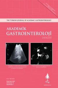
A 44-year-old man visited our outpatient clinic for a routine checkup with no complaint. He was not using any medications or alcohol. His vital signs and physical examination were normal. Liver enzymes were all within normal range, but cholestatic enzymes were mildly elevated: alkaline phosphatase: 124 (30-120 U/L), gamma-glutamyl transferase: 81 (0-55 U/L), and total bilirubin: 1.10 (0.3-1.2 mg/dL). Hepatobiliary ultrasonography (US) revealed a liver of slightly elevated size (16 cm) with normal structure and parenchymal echogenicity. Intra- and extra-hepatic bile ducts were not dilated. US did not visualize the gallbladder clearly (Figure 1). The patient was anxious when he learned that US showed an invisible gallbladder. Two months later, he visited our outpatient clinic again. He was still anxious. We learned that he sought medical attention at other hospitals, and repeated US revealed an invisible gallbladder, but doctors suspected a contracted or porcelain gallbladder. Computer tomography was performed, which confirmed the absence of a visible gallbladder with normal intra- and extra-hepatic bile ducts similar to suspected contracted or porcelain gallbladder. We performed magnetic resonance cholangiopancreatography (MRCP), which revealed normal intra- and extra-hepatic bile ducts and pancreatic duct with no dilation or filling defect. The gallbladder was not detected (Figure 2).
Gallbladder agenesis (GA) is a rare congenital anomaly of the biliary system with an estimated incidence of 13-65 individuals per 100 000 cases (1). Three groups of GA were identified in 1988 (2): asymptomatic (35%), which comprised individuals diagnosed at autopsy or laparotomy; symptomatic (50%), whose symptoms were chronic right upper quadrant pain (90%), dyspepsia (30%), nausea/vomiting (66%), fatty food intolerance (37%), and jaundice (35%); and foetal anomalies (15%), which indicated congenital anomalies incompatible with life. The most common clinical presentation is right upper quadrant pain mimicking biliary colic. Most cases of symptomatic GA are scheduled for surgery with a misdiagnosis of cholecystitis or choledocholithiasis. If the diagnosis of GA is made intraoperatively, patients often are exposed to complications from prolonged exploration. Aborting the procedure rather than complete further exploration is recommended (3). US is the most sensitive technique for imaging the biliary system. Most cases of GA have been reported as “contracted/fibrotic gallbladder” on ultrasound. The main method to establish GA is MRCP, which is a noninvasive and well-demonstrated imaging method in the evaluation of the biliary tract. It can also demonstrate an excluded and/or ectopic gallbladder (4,5).
MRCP enables the correct diagnosis to be made by a noninvasive examination, thereby suppressing our patient’s anxiety. The patient was informed about his congenital malformation to avoid possible unnecessary surgical exploration in the future. GA should be considered whenever the gallbladder is not visualized in routine imaging methods in patients with or without symptoms to reduce misdiagnosis and unnecessary surgeries.
Anahtar Kelimeler
Kaynakça
- 1. Kasi PM, Ramirez R, Rogal SS, Littleton K, Fasanella KE. Gallbladder agenesis. Case Rep Gastroenterol 2011;5:654-62. 2. Bennion RS, Thompson JE, Tompkins RK. Agenesis of gallbladder without extrahepatic biliary atresia. Arch Surg 1988;123:1257-60. 3. Balakrishnan S, Singhal T, Grandy-Smith S, El-Hasani S. Agenesis of the gallbladder: lessons to learn. JSLS 2006;10:517-9. 4. Vanbeckevoort D, Van LH, Ponette E, et al. Imaging of gallbladder and biliary tract before laparoscopic cholecystectomy: comparison of intravenous cholangiography and the combined use of HASTE and single-shot RAREMR imaging. J Belge Radiol 1997;80:6-8. 5. Adusumilli S, Siegelman ES. MR imaging of the gallbladder. Magn Reson Imaging Clin N Am 2002;10:165-84.
Ayrıntılar
| Birincil Dil | İngilizce |
|---|---|
| Konular | Sağlık Kurumları Yönetimi |
| Bölüm | Makaleler |
| Yazarlar | |
| Yayımlanma Tarihi | 29 Nisan 2019 |
| Yayımlandığı Sayı | Yıl 2020 Cilt: 19 Sayı: 1 |
test-5